 Home
Home
|
Table of Content - Volume 17 Issue 3 - March 2021
Study of lipid peroxidation and antioxidant activity in stored blood
Chhaya Keny1, Sucheta Dandekar2*, Jayashree Sharma3
1Technical Supervisor, 3Professor & HOD, Department of Transfusion Med., Kem Hospital, Parel, Mumbai, INDIA. 2Professor, Department of Biochemistry, Era University, Era's Lucknow Medical College, Uttar Pradesh, INDIA. {Adjunct Faculty, Manipal Aca of Health Education, INDIA.} Email: chhayakeny.ck@gmail.com, sucheta.dandekar@gmail.com, jayashreesharma@kem.edu
Abstract Background: Red blood cell is one of the important blood products that can be stored for 35-42 days in blood bank. The recipients are the patients, who need blood for increasing their oxygen carrying capacity. Red blood cells carry oxygen and transfer it to all body cells, but with continuous exposure to the oxidative stress. The red blood cells stored over time face changes in physical, biochemical and immunological specifications called RBC storage lesion. Oxidative damage is the most important factor causing RBC storage lesion which is caused by free radicals and can affect quality of red blood cellsor RBC. Free radicals can damage RBC by lipid and protein oxidation. This study is aimed to assess effects of lipid peroxidation and determine the levels of enzymatic antioxidants that act as protective safeguards. Material and methods: The present study was observational study carried out in healthy blood donors at KEM hospital, Mumbai. Thirty healthy donors, who were fulfilling the inclusion and exclusion criteria were selected. Properly stored blood samples at 40C were processed for estimation of hemoglobin, levels of lipid peroxidation and some enzymatic antioxidant activity. Enzyme levels estimation was carried out at every 7 days interval. Results: Malondialdehyde (MDA) is an indirect marker of lipid peroxidation that can modify proteins. Overall increase in MDA levels in the study indicates that lipid peroxidation in red cells has occurred to some extent from day 1 to the last day of the preservation. The study established positive correlation between lipid peroxidation and superoxide dismutase (SOD). On day 8, day 15 and day 22, lipid peroxidation was found to be positively correlated with SOD. But on Day 30, there was negative correlation between lipid peroxidation and SOD. On the whole, there was positive correlation between lipid peroxidation and SOD and it was statistically significant. Methemoglobin levels in stored blood were increased over a period of 30 days. Conclusion: From the present study, it can be concluded that though storage of blood can cause increase in lipid peroxidation, the antioxidant enzymatic machinery of the system comes into play adequately to circumvent the damage done. So, blood if stored properly can be safely used for transfusion till its expiry. Key Words: antioxidant, enzymes, superoxide, dismutase, lipid.
INTRODUCTION It is only since the last century that the lifesaving potential of blood and its components has been truly known and hence the importance of blood transfusion1. The first successful human blood transfusion took place in 1665 by Richard Lower2. Blood collected from the donor has to be stored under same physiological conditions as in vivo until it is transfused to the recipient, to maintain viability and activity of red cells, i.e. to maintain the quality of blood. The first attempt to store the blood was made during the First World War. The basic physical and chemical conditions optimized for storage of blood include temperature, pH, the source of energy of red cells and the anticoagulant used. Even though such conditions are maintained, various parameters change during storage referred to as storage lesions. Storage lesions of Red cells A major requirement of blood transfusion therapy is the preservation of red cells in vitro so that these cells will function and survive normally when transfused to a recipient. When a blood is stored in a standard preservative solution under prper storage conditions, the red cells undergo a series of deteriorative changes over a period of time. These progressive changes are collectively referred to as the storage lesions. These changes include metabolic (those disrupting the intracellular machinery of red cells) and Structural changes (those affecting the integrity of cell membrane). The metabolic changes occur in Adenosine triphosphate, 2,3 diphosphoglycerate, blood glucose, methemoglobin, glutathione and certain enzyme levels.3,4 A potential source of injury is oxidation, red cells stored in plasma which are in contact with air, exposed occasionally to light and agitation and which contains enough metal to foster generation of hydroxyl and other free radicals.5 These attack and damage fatty acids and lipids of red cell membrane – referred to as lipid peroxidation. Lipid peroxidation continues up to the formation of Malondialdehyde (MDA).6 Malondialdehyde is an indirect marker of lipid peroxidation. MDA can modify proteins and together with the changes of membrane lipids during lipid peroxidation, may be the main cause of damage to erythrocyte membranes and subsequent hemolysis.7,8 In addition, the oxygen attached to hemoglobin can be an endogenous threat. Red cells usually contain several safe guards to prevent this potentially serious injury. These safeguards are the enzymes called as Antioxidants. Antioxidants (Free Radical Scavengers) Biological systems protect themselves against the hazards of free radicals and other activated oxygen derivatives by converting them either to oxygen by oxidation or to water by reduction. Such compounds are called antioxidants. The antioxidant defence system is composed of a broad spectrum of complex and simple compounds ranging from antioxidant enzymes, vitamins, amino acids and related organic compounds to transition type metals. These compounds have specific molecular configuration that can deactivate oxygen derived active sites.9 The enzymatic cellular defence mechanism is provided by enzymes like superoxide dismutase, glutathione peroxidase, catalase and other related enzymes of glutathione such as glutathione reductase and glutathione synthetase. Various mechanisms responsible for their antioxidant action include direct interaction with oxidant and oxidizing agents; scavenging of free radicals and singlet oxygen; separation of transition metals form specific site of action or reduction of hyperoxides etc.10, 11 Blood is permanently exposed to oxidative stress and therefore it has a high antioxidant capacity.12 In the stored blood of donors, many factors increasing the demands on the antioxidant capacity can be observed. Consequently, damage to erythrocytes by free radicals may occur. This can be manifested in different ways. Any decrease in the antioxidant defence capability of an oxygen consuming system or increase in reactive molecules in the system leads to oxidative stress. Thus, oxidative stress implies that there is a natural balance between free radicals and antioxidant defence and cells are damaged when the antioxidants are depleted or the radical formation is increased beyond the defence capacity of the cell. Malondialdehyde (MDA) may serve as a marker of lipoperoxidation13 while sufficient activity of enzyme Superoxide Dismutase (SOD) and Glutathione Peroxidase (GPx) protects the blood against oxidative damage.14 It is useful to control the alteration of total antioxidant status and lipid peroxidation in stored blood at the different days. There are only few isolated reports on the oxidative damage. Some investigators have studied stored blood related changes in lipid peroxidation and antioxidant system during storage of blood, but the results are controversial.15,16 The present work is an attempt to study in detail the oxidative damage in terms of lipid peroxidation and some antioxidant enzymes which may develop during preservation.
MATERIALS AND METHODS The present study was carried out in the department of Biochemistry of KEM Hospital. The study commenced after obtaining the sanction of the hospital Ethics committee. The blood samples for the present study were collected from healthy voluntary blood donors. In this case, the study is not based upon clinical investigations; hence patients with any clinical disorders are not involved in this study. 30 healthy donors who were willing to donate their blood for this study purpose were selected by applying inclusion and exclusion criteria. Nature, objectives and benefits of the study were explained to each donor. Informed consent was obtained prior to entry into the study.
The blood was preserved at 40C±20C in citrate phosphate dextrose adenine (CPDA) preservative in cold room of blood bank for thirty days. Aliquots were drawn from the bag on the 1st day, 8th day, 15th day, 22nd day and 30th day of storage. All the under mentioned parameters were studied in each of 30 bags. Study Plan Volunteer’s blood was withdrawn after routine selection procedures (by confirming negative result for HIV,HBsAg, and VDRL tests). Following laboratory investigations were carried out;
The investigations were carried out with red cell membrane that was isolated by the method of hypnotic lysis of the red cells as suggested by Dodge.
The present work was undertaken with a view to study oxidative stress and enzymatic oxidants during blood preservation at 40C. All the statistical calculations namely mean, standard deviation, students paired and unpaired test ‘t’ test and correlation of coefficient were done using SPSS 10.01. Unit definition: Superoxide dismutase (SOD): 1 unit of SOD is the amount of required to cause 50% inhibition of pyragallol antioxidation per minute.
RESULTS Analysis of the samples at 7 days interval in the subjects is presented below;
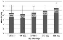
Table 1: Extent of Lipid peroxidation occurred during storage
Day1vs Day 8- p > 0.05 NS; Day1vs Day 15- p > 0.05 NS; Day1vs Day 22- p < 0.05*, Day1vs Day 30- p < 0.0)1*** Extent of lipid peroxidation measured on all 30 days by the amount of malondialdehyde (MDA) is given above.
Graph 1: Extent of Lipid peroxidation occurred during storage Table 2: Values of Superoxide dismutase over a period of 30 days
Day 1vs Day 8- p > 0.05* ; Day 1vs Day 15- p < 0.001*** ; Day 1vs Day 22- p < 0.001***, Day 1vs Day 30- p < 0.001*** It can be observed from above table 2, increasing levels of superoxide dismutase in 30-day measurement.
Table 3: Comparative Values of MDA and Superoxide dismutase
Day 1vs Day 8- p > 0.05* ; Day 1vs Day 15- p < 0.001*** ; Day 1vs Day 22- p < 0.001***, Day 1vs Day 30- p < 0.001*** Table 3 shows an increased superoxide dismutase level over a storage period of 30 days from day 15 onwards. This increase in superoxide dismutase level is statistically significant.
Graph 2: Values of Superoxide dismutase over a period of 30 days
Table 4: Comparative Values of MDA and Methemoglobin
Day 1vs Day 8- p < 0.01*; Day 1vs Day 15- p < 0.001***; Day 1vs Day 22- p < 0.001***, Day 1vs Day 30- p < 0.001*** Based on the data presented in Table 4, it can be observed that Methemoglobin levels in stored blood were increased over a period of 30 days. The increase on day 8th, day 22nd and day 30th is statistically significant.
Table 5: Correlation coefficients between SOD and Methemoglobin
*Correlation Significant at the 0.05 level (2-tailed) Table 5 above summarizes correlation coefficient between lipid peroxidation and antioxidant – SOD over a period of 30 days.
DISCUSSION Apart from hematological major markers like cell counts of leukocytes, erythrocytes or platelets; hemoglobin, hematocrit, mean corpuscular value (MCV), Mean corpuscular hemoglobin concentration (MCHC); levels of glycolytic intermediates like 2, 3 diphosphoglycerate are indicators to assess the quality of stored blood for transfusion.22 Red cells are stored in plasma which is in contact with air, exposed occasionally to light and agitation and it contains enough metal to foster generation of hydroxyl and other free radicals. Free radicals disrupt the equilibrium of lipid membrane of red cells by damaging their major constituent molecules. Therefore, lipid peroxidation has been sued as an indicator of oxidative stress. It can be determined by measuring the concentration of low molecular weight enzymatic antioxidant like superoxide dismutase (SOD). In the present study, overall increase in MDA levels indicate that lipid peroxidation in red cells has occurred to some extent from day 1 to 30th day of preservation. Same observation has been reported by Aslan R et al.15 and Korgun D K et al.16 in their studies in blood donors. SOD specially catalyzes the dismutation of superoxide anion radical to hydrogen peroxide and oxygen. In the red cell, SOD is a soluble cuprozonic enzyme with a molecular weight of about 32,000 and acts as first defence against oxygen reaction products. Red cells are quiet rich in superoxide dismutase enzyme. SOD is one of the most important antioxidant enzymes in the red blood cell.23 Present study showed an increase in SOD levels over the period of preservation. Comparative analysis of MDA and SOD levels indicate gradual increase of both over the storage period of 30 days. SOD also showed positive correlation with lipid peroxidation, correlation coefficient Being 0.22 (p=0.057). Websters et al. 24 found that SOD activity was found to reduce with storage beyond 10 days. Further focus research with more volunteer samples will be useful in understanding the phenomenon. Methemoglobin is an inactive form of hemoglobin because it cannot transport oxygen. Study results have shown that Methemolgobin levels in stored blood were increased over a period of 30 days. The increase on day 8th, day 22nd and day 30th is statistically significant.
CONCLUSION In the present study, slight increase in MDA level over a period of storage was observed indicating small amount of lipid peroxidation of the red cell membrane during preservation. Antioxidant enzymes which act as safeguards were active throughout the preservation period. There was an increase in superoxide dismutase levels and that was statistically significant. Therefore, it can be concluded that though storage of blood can cause increase in lipid peroxidation, the antioxidant enzymatic machinery of the system comes into play adequately to counter the damaging effect and protect the red cells membrane.
REFERENCES
Tanaka KR, Valentine WN, Miwa S. Pyruvate kinase (PK) deficiency hereditary nonspherocytic hemolytic anemia. Blood. 1962;19(3):267-295.Shafer, A. W, Bortelt G R Phosphorylated carbohydrate intermediates of the human erythrocyte during storage in acid citrate dextrose. J. clin. Invest.1961;40,1185.
Maxwell, S.R.J. Prospects for the use of antioxidant therapies, Drugs 1995; 49 (3). 345-361Halliwell B Free radicals, reactive oxygen species and human disease: a critical evaluation with special reference to atherosclerosis. Br J Exp Pathol. 1989 Dec;70(6):737-57.Stephen A. Levine, Parris M. Kidd The biochemistry of Free radicals in Antioxidant Adaptation: Its Role in Free Radical Pathology, Allergy Researcher group 1994; 48.
Marklund SL and Marklund G. Involvement of the superoxide anion radical in the autoxidation of pyrogallol and a convenient assay for superoxide dismutase. Eur. J. Biochem.,1974; 47, 469.
Sarma PR. Red Cell Indices. In: Walker HK, Hall WD, Hurst JW, editors. Clinical Methods: The History, Physical, and Laboratory Examinations. 3rd edition. Boston: Butterworths; 1990. Chapter 152Fayez M. Hafez, Hoda M. A.Hassab, Zeinab E. Mourad and Hamdy E. Ascalany Red Blood Cells Superoxide Dismutase Activity in Iron Deficiency Anemia Alex J Pediatr, 1999;13(2),439-42Webster N.R. · Toothill C. Effects of Blood Storage on Red Cell Antioxidative Systems Acta Haematol 1986;75:30–33.
Policy for Articles with Open Access
|
|
 This work is licensed under a Creative Commons Attribution-NonCommercial 4.0 International License.