 Home
HomeOfficial Journals By StatPerson Publication
|
Table of Content - Volume 4 Issue 2 - November 2017
Serum lipid profile in oral submucous fibrosis - A case control study
Arati Ganiger1, K Mallikarjuna Swamy2*
{1Tutor, Department of Biochemistry} {2Assistant Professor, Department of ENT} KIMS, Koppal, Karnataka, INDIA. Email: coolarati123@gmail.com
Abstract Background: Oral Sub Mucous Fibrosis (OSMF) is a chronic, debilitating disease characterised by juxtaepithelial fibrosis of the oral cavity. It is a precancerous condition a generalized pathological state of the oral mucosa associated with a significantly increased risk of oral cancer. Lipids are major cell membrane components. The changes in serum lipid levels have long been associated with cancerous and precancerous conditions. So this study is aimed to evaluate the plasma lipid profile in OSMF patients. Objectives: The present study aimed to evaluate the alteration in serum lipid profile in OSMF and to compare them with healthy controls and to correlate the relationship between pathogenesis of OSMF and lipid profile. Materials and Methods: It is a case control study. The study included 50 diagnosed cases of OSMF and 50 matched healthy controls. Fasting venous blood of 3 ml was collected in both cases and controls and serum was separated. Fasting serum lipid profile including Total Cholesterol (TC), Very Low Density Lipoproteins (VLDL), Low Density Lipoproteins (LDL), High Density Lipoproteins (HDL) and Tri-Glycerides (TG) were measured using automated analyser. Statistical analysis was done using student 't' test. Pearson's correlation was performed to establish the relationship between study variables. Results: The plasma total cholesterol, TG, LDL, VLDL and HDL levels were significantly reduced in patients with OSMF as compared to the control group.(p<0.005) Conclusion: Our study indicates that there is an inverse relationship between OSMF and serum lipid profile. Decrease in the lipid levels may be considered as a valuable biochemical marker in the early diagnosis and prognosis of oral malignancy. Key Words: OSMF, TC, HDL, VLDL, premalignant, Oral cancer, biochemical marker.
Oral submucous fibrosis (OSMF) is a chronic, debilitating disease characterised by juxtaepithelial fibrosis of the oral cavity1. It is a precancerous condition-a generalized pathological state of the oral mucosa associated with a significantly increased risk of cancer2 according to world health organization (WHO).Although occasionally preceded by, or associated with formation of vesicles, it is always associated with a juxtaepithelial inflammatory reaction followed by fibro elastic change of the lamina propria and epithelial atrophy that leads to stiffness of the oral mucosa and causes trismus and an inability to eat3. The predominant age group affected is 20-40 years though it can occur in any age group. Compared to traditional betel quid, gutkha chewing tends to begin at a younger age and has a shorter time to the development of disease, so cases of oral submucous fibrosis have been seen in as young as 11 years of age. OSMF is believed to be multifactorial though pathogenesis is not fully understood. Factors include areca nut chewing, ingestion of chillies, genetic and immunologic processes, nutritional deficiencies and other factors. Malnutrition, iron deficiency anemia, vitamin B complex deficiency are promoting factors that derange the repair of the inflamed oral mucosa, leading to defective healing and resultant scarring4. Tilakaratne et al reported that areca nut is the main etiological factor for OSMF5. Betel quid chewing is seen almost exclusively in the Indian subcontinent, South East Asia and Western Pacific. Excessive use of areca nut may cause fibrosis due to increased synthesis of collagen, and induce the production of free radicals and reactive oxygen species, which are responsible for high rate of oxidation/peroxidation of polyunsaturated fatty acids which affect essential constituents of cell membrane and may involve in tumorogenesis6,7,8. Because of the lipid peroxidation, there is a greater utilization of lipids for new membrane biogenesis. Cells fulfil these requirements either from circulation, by synthesis through the metabolism or from degradation of major lipoprotein fractions like VLDL, LDL or HDL7. Studies by various researchers have reported an association of plasma/serum lipids and lipoproteins with different cancers, as cholesterol is essential for maintenance of structural and functional integrity of all biological membranes. The present study aimed
MATERIALS AND METHODS This was a case control study. The study was carried out on 50 cases of clinically diagnosed OSMF in the age group of 20-50 years, attending the otorhinolaryngology (ENT) outpatient department (OPD), KVG Medical College, Sullia. Fifty (50) age and sex matched healthy subjects were taken as controls. The study was conducted over a period of one year from May 2016 to May 2017. Ethical clearance was obtained from the institute’s ethical clearance committee. Informed consent was taken from the cases and controls after explaining the procedure. The fifty OSMF patients were clinically examined and diagnosed. Later they were confirmed histopathologically following punch biopsy. Exclusion Criteria
Biochemical Analysis A fasting blood sample of 3 ml venous blood was collected under aseptic precautions in a plain vial. It was allowed to clot and serum was separated by centrifugation. Lipid profile was analysed using automated analyser by following methods
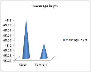
Statistical Methodology: Data was expressed in terms of mean ± SD. Chi- square test was applied to estimate the difference between the two groups of population. Unpaired ‘t’-test was used to study the changes in serum lipid levels between the study groups. Pearson correlation was performed to establish the relationship between study variables. p value <0.05 was considered statistically significant. RESULTS This was a comparative case control study conducted on 50 cases of OSMF (n=50) and 50 age and sex matched healthy controls (n=50). Serum lipid levels were estimated and analyzed in both cases and controls. The results were expressed as mean ± standard deviation. The age distribution of cases and controls is depicted in Table 1, Figure 1. The mean age (in years) of cases was 45.5±11.7 years and that of controls was 45.2 ±10.3 years and was not significant. (p>0.05). Table 2 shows the gender distribution. Out of 50 cases of OSMF, 30(60%) were males and 20 (40%) were females. Out of 50 controls, 30(60 %) were males and 20 (40%) were females and it was not statistically significant (p=0.38). In general, males were more commonly affected than females. The incidence of OSMF was most commonly seen between 2nd and 4th decade. The mean serum Total Cholesterol, serum HDL, serum LDL, serum VLDL and serum TG levels in OSMF cases and control group are shown in Table 3. A statistically significant reduction [p<0.001] was noted between the control group and OSMF cases for all lipid parameters. (Table 3, Figure 2).
Table 1: Age distribution of cases and controls
OSMF=Oral Submucuous Fibrosis
Table 2: Gender distribution of cases and controls
X2= 4.165 p=0.38, Not significant Table 3: Comparison of serum lipid levels between controls and OSMF
OSMF-Oral Submucous Fibrosis, TC-Total Cholesterol, LDL-Low Density Lipoprotein, TG-Triglyceride, VLDL-Very Low Density Lipoprotein
Figure 1: Mean age of cases and controls Figure 2: Comparison of serum lipid levels in between OSMF cases and controls TC: Total Cholesterol, TG: Triglyceride, HDL: High Density Lipoprotein, VLDL: Very Low Density Lipoprotein, LDL: Low Density Lipoprotein
DISCUSSION Lipids are major cell membrane components essential for various biological functions including cell growth and division of normal and malignant tissues. The changes in lipid profile have long been associated with cancerous and precancerous conditions because lipids play a key role in maintenance of cell integrity9. As lipids may play a role in precancer and cancer, this study was aimed to estimate serum lipid profile in oral submucous fibrosis groups and to compare the values with those values from control groups. Our study showed a decreased levels of serum lipids in cases as compared to controls. The decrease in serum lipids in patients with OSMF could be due to
The habit of tobacco consumption is a known etiologic factor for development of oral precancerous diseases and head/neck cancer10,11. Cholesterol which is an amphipathic lipid is an essential structural component of all cell membranes and of the outer layer of plasma lipoproteins. It is present either as free cholesterol or combined with a long-chain fatty acid, as cholesterylester in tissues and in plasma lipoprotein. It is synthesized from acetylCoA in many tissues and is ultimately eliminated as cholesterol or bile salts from the body. In the circulation, lipoprotein transports free cholesterol and it readily equilibrates cholesterol in other lipoproteins and in membranes12-15. Free radicals and reactive oxygen species are generated from tobacco carcinogens which are responsible for high rate of oxidation / peroxidation of polyunsaturated fatty acids. It results in greater utilization of lipids including total cholesterol, lipoproteins and triglycerides for new membrane biogenesis. Cells fulfil these requirements either from circulation, or by synthesis through metabolism or from degradation of major lipoprotein fractions like VLDL, LDL, and HDL. Earlier studies have shown alteration of plasma lipid profiles in head and neck and other cancers13-15. In the present study, a significant decrease in serum total cholesterol, HDL, LDL, VLDL and TG was observed in oral OSMF patients as compared to the controls. The results in our study add to the evidence of an inverse relationship between serum lipid profile and oral submucous fibrosis. The decrease in total cholesterol in patients with OSMF could be due to the excessive use of areca nut and also due to rapidly dividing cells in malignancy. In areca nut, the major alkaloid arecoline undergoes nitrosation and gives rise to N- nitrosamine, which might have cytotoxic effect on the cells. This leads to lipid peroxidation by reactive free radicals generation and also leads to greater utilization of lipids as explained above16,17. In our study, the decrease in total cholesterol levels may be a useful indicator reflecting initial changes occurring in precancerous and neoplastic conditions. The evidence suggests that in precancerous conditions like OSMF, cells are able to remetabolize lipids for their growth and to generate phospholipids membranes. Recent progress in molecular biology will assist researchers in the near future to identify the genes and enzymes of lipid metabolic pathways.
CONCLUSION Our study shows that there is an inverse relationship between serum lipid profile and OSMF. The change in plasma lipid levels may be used as a diagnostic or prognostic biochemical indicator for early diagnosis of oral premalignant and malignant conditions. However, a detailed study on large sample size and on role of cholesterol in neoplasia should be carried out for better understanding of this inverse relationship of serum lipid profiles and oral pre malignant and malignant conditions. From the present study, the lower serum lipid levels may have a diagnostic or prognostic role in the early diagnosis of Oral premalignant and malignant lesions.. Low levels of cholesterol could be due to the rapidly dividing cells in premaliganincies and malignancies.
REFERENCES
|

 This work is licensed under a Creative Commons Attribution-NonCommercial 4.0 International License.