 Home
Home
Official Journals By StatPerson Publication
|
Table of Content - Volume 11 Issue 3 - September 2019
Concurrent tubal ectopic pregnancy with cesarean scar ectopic pregnancy: World's first case report
Ammbalal Gurram1*, Garcha2, Shital Takpire3, Vivek Kulkarni4, S Kurhade5, Anjali Ghadge Gurram6
{1Senior Consultant, Gyanecologist & Laparoscopic Surgeon} {2Sr Consultant, Department of Anaesthesiology} {3Department of Anesthesiology} {4,5Lap Surgeon} Dr D Y Patil Medical College & Hospital, Pimpri, Pune, Maharashtra, INDIA. 6Consultant, Ashwini Nursing Home, Pune, Maharashtra, INDIA. Email: ammbalalgurram@gmail.com
Abstract Cesarean Scar Ectopic is one of the rarest of all ectopic pregnancies. Concurrent tubal ectopic pregnancy with cesarean scar ectopic pregnancy is the first case to be reported. The incidence of cesarean scar ectopic pregnancy is on increase due to increase in number of cesarean Deliveries and early diagnosis by ultrasound. Tubal ectopic pregnancy is the most common type of ectopic pregnancy and incidence of tubal ectopic pregnancy is also increasing due to increase in incidence of tubal disease. Early diagnosis and management is extremely important as delay in diagnosis and management of ectopic pregnancy can lead to maternal mortality and morbidity. MRI scan is a useful tool when ultrasound is inconclusive before proceeding to management or surgery. We are reporting the World's First Case of Concurrent tubal ectopic pregnancy with Cesarean scar ectopic pregnancy. A 37 year old client with G3 P1 L1 A1, had a history of previous abortion and a Cesarean section was diagnosed on Ultrasound as concurrent tubal ectopic pregnancy with Cesarean scar ectopic pregnancy. Initially managed with Medical management with Methotrexate. The tubal ectopic showed resolving but Cesarean scar ectopic pregnancy was not resolving with medical management. Decision Laparoscopic management of ectopic pregnancy was taken. She underwent Laparoscopy with hysteroscopy. Histopathological report confirmed tubal ectopic pregnancy with Cesarean scar ectopic pregnancy. As ectopic pregnancy is a potentially life threatening condition early diagnosis and management can avoid the complications and mortality and morbidity. Key Words: Tubal ectopic pregnancy, Cesarean scar ectopic pregnancy.
INTRODUCTION Ectopic pregnancy is a potentially life-threatening condition occurring in 1-2 % of all pregnancies. The most common ectopic implantation site is the fallopian tube, though 10 % of ectopic pregnancies implant in the cervix, ovary, myometrium, interstitial portion of the fallopian tube, abdominal cavity or within a cesarean section scar. Cesarean scar pregnancy (CSP) is a rare entity and can cause serious complications.1The first case of CSP was reported by Larsen and Solomon in 1978.2 There is a rising trend in the number of cases being reported possibly due to the increasing prevalence of cesarean sections. Increasing the use of imaging studies such as ultrasonography and magnetic resonance imaging (MRI) helps in detecting these cases. Early diagnosis would help avoid complications such as scar rupture and excessive hemorrhage, which may require a hysterectomy. This can endanger the woman’s life and also affect future fertility.3Patients who are vitally stable have more treatment options including conservative management. Hence, obstetricians/gynecologists and radiologists must be highly vigilant of this potentially fatal complication.4
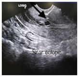
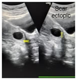
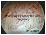
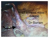
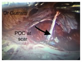
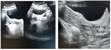
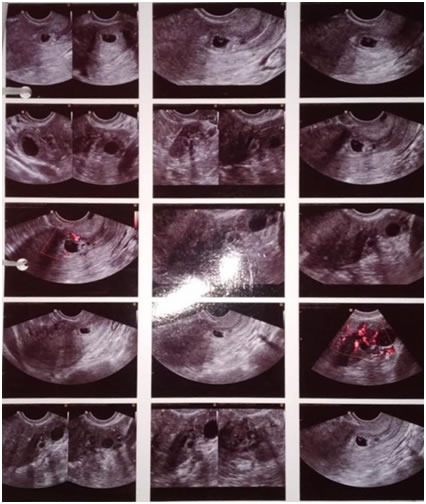
CASE REPORT A 37 year old female came to Out Patient Department with history of 2 months amenorrhea and complained of pain abdomen. Her Obstetric History was G3P1L1A1. Her first delivery was Cesarean section done for fetal distress 9 years ago, had a living male child. Her second pregnancy had abortion, Medical Termination of Pregnancy. On examination the general condition was good, her vitals were stable, systemic examination normal, on per abdomen examination there was mild tenderness in left illiac fossa, per speculum examination cervix healthy, no discharge and per vaginum examination showed uterus bulky, tenderness in left fornix while right fornix free and not having any tenderness. Routine investigations were carried out and came out to be in normal range. Serum B HcG was 5644 miu/ml initially and was 7888 miu/ml after 48hrs which was less than double suggestive of ectopic pregnancy. Transvaginal ultrasound was carried out showed a small gestational sac in left adnexa in close proximity of left ovary with fairly maintained fat planes. Gestational Sac 6.2mm = < 5 weeks. It also showed another gestational sac 12.8mm low lying in uterine cavity in close proximity to Cesarean Scar. Adjacent anterior myometrial wall showed thinning upto 6.4mm. Gestational Sac= 5weeks 3 days yolk sac present. Both sac didn't have fetal pole and had no cardiac activity. On colour doppler examination showed excessive vascularity suggestive of concurrent tubal ectopic and Cesarean scar ectopic pregnancies. As the patient was suitable for medical management of ectopic pregnancy, she was given Injection Methotrexate 50mg intramuscularly and BHcG was monitored every 3-4 days. After 4 weeks B HcG was 204 miu/ ml. Transvaginal ultrasound was done showed Cesarean scar ectopic not resolved and ? tubal abortion as tubal ectopic was not seen clearly. As patient was heamodynamicaly stable close monitoring of patient done. After 2 weeks Ultrasound was done which showed Cesarean scar ectopic pregnancy was not resolving so decision of surgical management was taken. Diagnostic Hysteroscopy was done to rule out intrauterine pregnancy. No intrauterine pregnancy was seen. Laparoscopy was done showed left fallopian tube ward congested and cynosed. Laparoscopic left salpingectomy done. Approximately 200ml of blood clots removed from pouch of Douglas suggestive of tubal abortion. Then bladder fold dissected and bladder separated. The Cesarean scar excised gush of fluid came and products of conception seen clearly and removed completely. Suturing was done Laparoscopically. Hemostasis confirmed. Procedure done uneventfully. Patient tolerated the procedure well. Left fallopian tube and excised Cesarean scar ectopic products of conception sent for Histopathological examination which confirmed concurrent tubal ectopic pregnancy with Cesarean scar ectopic pregnancy.
Figure 1: first scan showing scar ectopic scan - scar ectopic Figure 2: Hysteroscopy pic showing no pregnancy Left adnexa – ectopic scar ectopic POC Inside This is a Case Report of World's First Case of Concurrent Tubal Ectopic Pregnancy with Cesarean Scar Ectopic Pregnancy. Figure 4: combination of all images - tubal ectopic and scar ectopic
DISCUSSION Cesarean scar ectopic pregnancy is the rarest of all ectopic pregnancies and tubal ectopic pregnancy is the most common type of ectopic pregnancy. But concurrent tubal ectopic pregnancy with Cesarean scar ectopic pregnancy has not been reported yet. This is a Case Report of World's First Case of Concurrent Tubal Ectopic Pregnancy with Cesarean Scar Ectopic Pregnancy. An ectopic pregnancy occurs when a fertilized ovum implants outside the normal uterine cavity. It is the most important cause of maternal mortality and morbidity in the first trimester. Risk factors like previous ectopic pregnancy, tubal corrective surgery, tubal sterilization, intrauterine devices, documented tubal pathology, infertility, assisted reproductive techniques, PID, smoking, prior abortions, multiple sexual partners and prior delivery have been implicated in the development of the ectopic pregnancy. A knowledge of the associated risk factors helps identify women at higher risk in order to facilitate early and more accurate diagnosis. Most risk factors are associated with risks of prior damage to the Fallopian tube. These factors include any previous pelvic or abdominal surgery, and pelvic infection. Chlamydia trachomatis has been linked to 30-50% of all ectopic pregnancies. Early diagnosis reduces the risk of tubal rupture and allows more conservative medical treatments to be employed. The exact cause of CSP is still not clear. There is an early invasion of the myometrium and it is presumed that this occurs through a microscopic tract in the cesarean section scar tissue. 5The incidence has been reported to be 1:1 800 to 1:2 200 pregnancies. 5,6In CSP, the gestational sac gets embedded within the fibrous tissue of the previous cesarean section scar.7 The gestational age at diagnosis ranged from five to 12.4 weeks (mean 7.5 ± 2.5 weeks) and the time interval between the last cesarean and the CSP was six months to 12 years.8 There are many risk factors implicated in the development of CSP. These include the number of cesarean sections, the time interval between the previous cesarean section and the subsequent pregnancy, and the indications for the previous cesarean section, but it is not clear whether these factors are directly related to CSP. 8On review of the various case reports, it was noted that CSP were incidental ultrasonography finding in an asymptomatic woman while some present with mild painless vaginal bleeding. In a lesser percentage of patients, it was accompanied with mild to moderate abdominal pain. The uterus may be tender during examination if the CSP is in the process of rupture. A patient with a ruptured CSP may present in a state of collapse or hemodynamically unstable.8 Transvaginal sonography remains to be an important tool in diagnosing CSP and could soon be the gold standard for the diagnosis of scar implantation.5 Diagnostic criteria are as follows: ■ An empty uterine cavity and an empty cervical canal. ■ A gestational sac in the anterior part of the uterine isthmus. ■ An absence of healthy myometrium between the bladder and gestational sac. 5 ■ Circular blood flow surrounding the sac must also be clearly visible.9-11 CSPs were noted to be well perfused on Doppler examination. 5 Usually a transvaginal scan combined with color flow Doppler can be a reliable tool in diagnosing a CSP. MRI may be used as an adjunct to ultrasound scan. As it is a rare condition, there are no specific guidelines available for the management of CSP. The main aim of treatment of CSP is to prevent massive blood loss and conserve the uterus to maintain future fertility, women’s health, and quality of life.12 Management plans are made based on the gestational age, hCG levels, and presence of cardiac activity. Management may be either medical or surgical. Various treatment options include dilatation and curettage and excision of trophoblastic tissues using laparotomy or laparoscopy, local and/or systemic MTX administration, bilateral hypogastric artery ligation, associated with dilatation and evacuation under laparoscopic guidance, and selective uterine artery embolization (UAE) in combination with curettage and/or MTX injections. 1, 13 Conservative management has been considered when there is a silent miscarriage in the scar in very early pregnancies and beta-hCG values are closely monitored. 14, 15 Conservative medical management has been recommended for women who are asymptomatic and hemodynamically stable with unruptured CSP of less than eight weeks gestation and a sonographic finding of myometrial thickness less than 2 mm between the CSP and the bladder.7 Use of MTX locally, systemically, or combined is recommended as the first line of treatment for CSPs. Laparoscopy for CSP treatment is recommended for cases that are hemodynamically stable and have a deeply implanted gestational sac growing towards the abdominal cavity and bladder. The surgery may be converted to a laparotomy if there is difficulty in achieving hemostasis.12 Surgical treatment is recommended when medical management fails or when a patient is hemodynamically unstable. Observation of a large amount of free fluid in the pelvic cavity on ultrasound scan will require surgery. Internal iliac ligation with partial lesion resection and uterine neoplasty has significant advantages in the surgical treatment of CSPs.14Huanxiao et al,27 reported 40 cases of CSP and recommended a transvaginal hysterotomy approach for removal of the ectopic tissue and repair of the uterine defect. They found the approach to be safe, cost effective, shorter hospital stay, and less postoperative pain and blood loss.17 Surgical treatment has the advantage that the gestationalanl mass can be removed and the defect can be repaired simultaneously, however, none of the treatment modalities can guarantee uterine integrity.15Jurkovic et al,5 recommended surgical repair of the scar either as a primary treatment or as a secondary operation after the initial treatment in women who desire further pregnancies. This could decrease the risk of recurrence of CSP. Once the gestational mass is surgically excised, it has been noted that hCG returns to normal much more quickly within one to two weeks. Dehiscence and repeat scar pregnancy have been reported following successful medical treatment with local MTX injections.18 Laparaoscopy and medical therapy have now emerged as the widely used therapeutic modalities with great succession in terms of reduced morbidity, shorter hospital stay and conservation of fertility.However choice depends upon early identification of ectopic pregnancy and stable condition of patients. Morbidity included anemia, blood transfusion and wound infection. By reducing and identifying the risk factors and catching the patients at the earliest it is possible to improve the prognosis so far as morbidity, mortality, and fertility are concerned. .
CONCLUSION The incidence if ectopic pregnancies are on the rise. All the cases were diagnosed with a high index of clinical suspicion and the USG findings added to the diagnosis. Though the recent trend in the management of ectopic pregnancy is the use of a conservative surgical or medical line of management, salpingectomy Surgical management is needed in many cases. CSP is a life threatening condition and the incidence is rising due to the increasing incidence of cesarean sections. It can have catastrophic complications affecting maternal morbidity and loss of future fertility. The liberal use of transvaginal ultrasound to assess early pregnancies helps early diagnosis and planning of the management. If the condition is not diagnosed, a simple gynecological procedure such as a dilatation and curettage may end up with massive hemorrhage and unexpected complications. The present day clinician should be aware of such a condition and have a high index of suspicion. Every pregnant woman with a past history of a cesarean section should have a careful ultrasonographic assessment of the previous scar. 16 As there are no evidence based recommendations available, clinicians will have to depend on the available case reports and counsel the women accordingly on the various treatment options available to make an informed choice. Consultants should be involved in patient counseling and planning the further management of such cases.
REFERENCES
|
|
 This work is licensed under a Creative Commons Attribution-NonCommercial 4.0 International License.