 Home
Home `
`
|
Table of Content - Volume 19 Issue 2- August 2021
Post shoulder joint manipulation sequelae -brachial plexus palsy - A rare case study report
P Udhayasingh1, Rajakumar2*
1Department of PMR, Tirunelveli, Medical College, Tirunelveli, Tamil Nadu, INDIA 2Department of PMR, Madras Medical College, Chennai, Tamil Nadu, INDIA
Abstract Frozen shoulder (FS), also known as adhesive capsulitis, is a common cause of a painful shoulder with restricted motion. The prevalence of frozen shoulder is estimated to affect 2–5% of the population, and affects men more than women.1,2 The peak incidence is observed between 40 and 60 years.1,2 Number of treatments options are available for frozen shoulder including drugs, physical therapy, Intra articular steroid injections, Hydro-dilatation, Supra scapular nerve block and manipulation under regional/general anaesthesia.3 Manipulation under anaesthesia (MUA) is a method wherein fibrosed capsulo-ligament complex of shoulder, which is a hindrance in regaining ROM, is forcibly broken by manoeuvring the shoulder across the ROM under anaesthesia. Soft tissue damage, shoulder dislocation, humerus fracture with rare involvement of nerve damage(brachial plexus) may occur after shoulder manipulation. We report case study report of brachial plexus palsy developed as post shoulder joint manipulation done for frozen shoulder. Key Word: brachial plexus.
CASE STUDY 60 years old male, known diabetic, presented with Right side shoulder pain with arm weakness for the past 4 months which followed manipulation of shoulder joint manipulation under general anaesthesia. Apparently he was normal ten months back. Later he developed pain and stiffness of shoulder for which he had physical therapy and drugs. Since there was no improvement, he underwent Right side shoulder manipulation 4 months back. Post manipulation aggravated the shoulder pain and minimal improvement in PROM, radiating pain from right side of neck to the arm developed after manipulation. Shoulder pain aggravated with weakness of right shoulder joint developed after shoulder joint manipulation. On examination, wasting and weakness of right side supraspinatus, infraspinatus and deltoid. AROM and PROM of right shoulder joint painful and restricted. other joints were normal. sensation, reflexes normal. left upper limb and both lower limbs normal. On local examination tenderness over supra clavicular region, acromio-clavicular joint and lateral aspect of shoulder joint noted.
SHOULDER JOINTS Figure 1: Right shoulder joint; Figure 2: Left shoulder joint
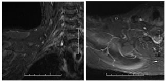
INVESTIGATIONS Xray shoulder joint normal on both sides prior to shoulder joint manipulation. Nerve conduction study: right supra scapular and axillary nerves CMAP amplitudes reduced. MRI brachial plexus: Mild thickening with hyper intensity signals were noted involving proximal division of right brachial plexus without obvious discontinuity. Partial tear of supraspinatus tendon on right side.
X-RAY SHOULDER JOINTS Figure 3:MR IMAGING OF RIGHT SHOULDER
PARTIAL TEAR OF SUPRASPINATUS TENDON AND ACROMIO-CLAVICULAR JOINT ARTHROPATHY Figure 4
BRACHIAL PLEXUS Mild thickening with hyper intense signals involving proximal divisions of right brachial plexus. Figure 5
MRI BRACHIAL PLEXUS Mild thickening with hyper intense signals involving the proximal divisions of right brachial plexus without any obvious discontinuity. {continuity is maintained}. Rest of the right brachial plexus appears unremarkable. No obvious evidence of pseudo meningocele is noted. RIGHT SIDE BRACHIAL PLEXUS - NO PSEUDO MENINGOCELE Figure 6: Conservative treatment included analgesic drugs, neuromodulators, B-vitamin complex and physiotherapy. Spontaneous recovery appeared at 14 months.
DISCUSSION Frozen shoulder is classically described as a benign condition consisting of three stages: painful freezing, adhesive or frozen, and the thawing or recovery phase. Many patients experienced persistent pain accompanied with limited range of motion (ROM). Krall et al.4 suggested that MUA is an effective method to improve function in patient with refractory FS in stage II, external rotation < 50% compared to opposite side and failure to respond to IA steroid infiltration.4 Literature reports an overall complication rate of 0.4%, and a re-intervention rate of 14%.4 Serious complications of MUA have been reported, such as a humeral shaft fracture, glenoid rim fracture, shoulder dislocation, brachial plexus traction injury or intra-articular damage to the cartilage or rotator cuff.5,6,7 MUA is performed under single shot interscalene brachial plexus block or if necessary general anesthesia. The scapula is indirectly stabilized by the supine position, a short lever arm and 90 degrees of elbow flexion is used to prevent fractures and brachial plexus traction injuries. Brachial plexus palsy can occur as a result of trauma, inflammation or malignancy. Manipulation of shoulder joint caused traction injury of mainly upper brachial plexus palsy due to forced pull by widening of shoulder and neck. Supraspinatus tendon partial tear may be due to shoulder manipulation. Nerves are vulnerable to injury from traction because the distance between the anchorage points of nerves in the upper limb is short especially for radial and musculocutaneous nerve. Axillary nerve is most frequently involved because it is relatively fixed as it travels through the quadrangular space and its course around the surgical neck of humerus. The use of a small lever arm and scapular stabilization is recommended to prevent fractures and brachial plexus traction injuries. 8,9 Birch treated three cases of brachial plexus injury post shoulder manipulation, one of which included a fracture of the proximal humerus.10 Diagnosis of brachial plexus injury is easy to miss as symptoms may be masked by pain due to bony injury or massive rotator cuff tear. Clinical suspicion is required, and in doubtful cases EMG and NCV must be done at 3-4 weeks post injury. Brachial plexus damage is the least observed complication following shoulder manipulation. Manipulation of shoulder joint causing brachial plexus traction injury mainly upper trunk makes this study a rare presentation.
REFERENCES
Policy for Articles with Open Access
|
|



 This work is licensed under a Creative Commons Attribution-NonCommercial 4.0 International License.