 Home
Home|
Table of Content Volume 3 Issue 1 - July 2017
Morphological study of lumbrical muscles in human cadaveric hands
Vrushali V Maindarkar1*, Santoshkumar A Dope2*, Pramod R Kulkarni3
1Assistant Professor, Department of Anatomy, MIMSRMC, Latur, Maharashtra, INDIA. 2Associate Professor, Department of Anatomy, GMC, Latur, Maharashtra, INDIA. 3Professor, Department of Anatomy, Dr. VMGMC, Solapur, Maharashtra, INDIA. Email: shiledar.vrushali@gmail.com, drdopesantosh@yahoo.co.in
Abstract The lumbrical muscles of hand, by producing flexion at metacarpo phalangeal (MCP) joints and extension at interphalangeal joints help in writing, stitching and other forms of precision work. Hence philosophically, it may be said that the actions of lumbrical muscles of hand are indices of civilization of a race.1 The variations in origin, insertion and nerve supply may cause carpal tunnel syndrome. Their hypertrophy may cause compression of radial and ulnar collateral arteries leading to chronic subischemia. Also they may cause surgical complications in hand surgeries. So this study has been undertaken to know about morphology and morphometry of lumbrical muscles in human hands.
INTRODUCTION The human hand is a prehensile organ. It is endowed with grasping and precision movements for skilled work and it acts as a chief tactile apparatus. This is contributed by a high degree of neuromuscular co-ordination and a larger cortical representation of the hand in sensory and motor cortex in human brain.2 Evolution of grasping ability of human being contributed by lumbrical muscles is attributed to the ecological context of such skills in the frogs. Hence human hand is revolution in evolution. The lumbricals of the upper limb are 4 small muscles resembling earthworms hence the name. They are numbered 1 to 4 from lateral to medial side. They arise from bare areas of tendons of flexor digitorum profundus (FDP) about the middle of the palm. The narrow tendon of insertion joins the radial margins of extensor expansion (EE) as distal wing tendons1 Normally 1st and 2nd lumbricals are unipennate. They arise from the radial sides of FDP tendons for index and middle fingers. They are supplied by branches from Median nerve. 3rd and 4th lumbricals are bipennate. They arise from adjacent sides of FDP tendons for middle, ring and little fingers. They are supplied by deep branch of ulnar nerve. Then they pass distally along the radial side of MCP joints anterior to deep transverse metacarpal ligament. The narrow tendon of insertion joins the EE of respective fingers as distal wing tendons. Through EE, they are inserted into dorsal surfaces of bases of middle and distal phalanges.
MATERIALS This study was conducted in the Department of Anatomy in an institutional Medical College, with 50 forearms and hands (25 Right and 25 left) of human embalmed cadavers of both sexes with the age range approximately between 45-65 years. 21 cadavers were male (42 hands) and 4 cadavers were female (8 hands). The study was carried out during the routine dissection for undergraduate medical students in the department of Anatomy. Material used for dissection was scalpel, blades, tooth and blunt forceps, scissor, scale, thread, marker pencil. METHODS As a guide for dissection, Cunningham’s manual of practical anatomy was used. The dissection was carried out as follows: A longitudinal incision was taken from the distal end of the flexor retinaculum, up to the level of the metacarpophalangeal joint of the middle finger. The skin, the superficial fascia, the deep fascia and the flexor retinaculum were dissected and reflected. Then, the tendons of the flexor digitorum superficialis and the flexor digitorum profundus were identified and reflected distally. The lumbrical muscles which were situated at the distal end of the flexor digitorum profundus tendons were carefully observed. The lumbrical muscles were followed to their tendons and traced up to their insertion.
OBSERVATIONS The following parameters were noted:
RESULTS Present study was carried out on 50 hands out of which 25 hands were of right side and 25 hands were of left side.
Table 1: Morphological comparison
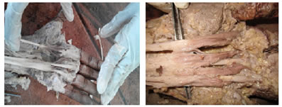
Right hand lumbricals- In present study, in 23 hands (92%) muscles were found normal morphologically, while in 2 hands (8%) 2nd lumbricals were bipennate originating from adjacent sides of tendons of flexor digitorum profundus (FDP) for index and middle finger. No misplaced insertions were found in any muscle. Left hand lumbricals- In present study, in 20 hands (80%) muscles were found normal morphologically. While in 4 hands (16%) 2nd lumbricals were bipennate originating from adjacent sides of tendons of FDP for index and middle finger. In 1 hand (4%) misplaced insertion in 4th lumbrical muscle was found at ulnar side of extensor expansion of ring finger instead of radial side of little finger. Figure 1: showing bipennate second lumbrical muscle Figure 2: showing all 4 lumbrical muscles DISCUSSION 1st lumbrical muscle Table 2: showing comparison of 1st lumbricals
In a study conducted by Ajmani [2001] in 68 hands, bifid 1st lumbrical was seen in 3 hands (4.5%) out of which 2 originating from flexor digitorum superficialis (FDS) tendon for index finger and 1 from palmar aspect of 2nd metacarpal bone. While in other 3 hands (4.5%) it was bipennate arising from adjacent sides of tendons of FDP and flexor pollicis longus. In a study done by Hosapatna et al [2013], bifid 1st lumbrical was found in 1 case out of 30 hands with an incidence of 3.3% and hypertrophied 1st lumbrical, in 1 hand (3.3%) out of 30. In a study conducted by Sawant et al [2013], 5 cases (5%) of accessory belly of 1st lumbrical were found out of 100 hands. In all cases the belly was originated from radial side of the tendon of FDP going to index finger. In the present study, 1st lumbricals in all 50 hands (100%) were present and found normal. They all were innervated by median nerve. 2nd lumbrical muscle Table 3: showing comparison among 2nd lumbricals
Ajmani [2001] found absence of 2nd lumbrical in both hands (3%) of a cadaver out of 68 hands. In 16 hands (24%) it was found to be bipennate. Joshi et al [2005] have found 2nd lumbrical bulkiest amongst all the lumbricals. They have also reported bipennate 2nd lumbricals in 32 hands (45%) out of 70 hands. It was also observed that the proximal attachment of 2nd lumbrical was extending to carpal tunnel compared to the rest of the lumbricals. Mutalik [2011] reports bipennate 2nd lumbrical in 2 cases (3.3%) out of 60 dissected hands. Fibers were arising from adjacent sides of FDP tendons for index and middle fingers. In a study done by Hosapatna et al [2013], bipennate 2nd lumbrical was found in 1 case (3.3%) out of 30 hands in which fibers were arising from adjacent sides of FDP tendons for index and middle fingers. Its pattern of insertion was normal. Sawant et al [2013] have noted 10 cases (10%) of bipennate 2nd lumbrical out of 100 hands, fibers arising from radial side of tendon of FDP for middle finger and ulnar side of FDP tendon for index finger. While in 7 hands (7%), it was found to be bifid originating from radial side of tendons of FDP and FDS muscles for index finger. Pattern of insertion was normal and there were no associated neurovascular variations. In a study done by Modasiya et al [2013], in 10 cases (12.5%) out of 80 hands, they found bipennate 2nd lumbrical. In the present study, in 2 right hands (8%) 2nd lumbricals were bipennate originating from adjacent sides of tendons of FDP for index and middle finger out of 25 right hands. While in 4 left hands (16%) of 2nd lumbricals were bipennate originating from adjacent sides of tendons of FDP for index and middle finger out of 25 left hands. 3rd lumbrical muscles Table 4: showing comparison of 3rd lumbricals
Where NS = nerve supply, ins = insertion, DTML =deep transverse metacarpal ligament, FDS = along flexor digitorum superficialis, mispl = misplaced St.John Brooks [1887] have reported variation in nerve supply of 3rd lumbrical in 12 hands out of 21 hands. He noticed a twig from median nerve as a sole nerve supply in 9 hands while dual nerve supply in remining 3 hands. In the hands with dual supply, the size of twig was inversely proportional to each other. Ajmani [2001] has reported 3rd lumbrical as the most variable lumbrical. He noted normal origin of all 68 hands but reported normal insertion in 32 hands (47%) only. The muscle was unipennate in 2 hands (3%). Split insertions were noted in 22 hands (32%). Insertion in base of first phalanx was noted in 10 hands (14%). Insertion into deep transverse metacarpal ligament was noted in 2 hands (3%) and along with FDS tendon in 2 hands (3%). Mutalik [2011] have reported split insertion of 3rd lumbrical in 7 cases (11.6%) out of 60 cases. She also noted misplaced insertion of it in 1 case (1.66%) where it was inserted into ulnar side of middle finger. Igiri et al [2011] have noted unipennate 3rd lumbrical muscle in 12 cases (18.75%) out of 64 hands. Parminder Kaur [2013] has reported variations in insertion rather than origin. It was seen that 14 hands (28%) out of 50 cases showed split insertions in which the tendon divided into 2 slips and inserted into ulnar side of middle finger and radial side of ring finger. It was also noted to have misplaced insertions in 2 hands (4%) in which it inserted into ulnar side of middle finger. Hosapatna et al [2013] reported absence of 3rd lumbrical in 1 hand (3.3%) out of 30 cases. Modasiya et al [2013] have reported split insertion of 3rd lumbrical in 12 hands (15%) of 80 cases. In the present study, 3rd lumbricals in all 50 hands (100%) were present and found normal. They all were innervated by deep branch of ulnar nerve. 4th lumbrical muscle Table 5: showing comparison of 4th lumbricals
Where mispl ins = misplaced insertion, Ajmani 2001 reported absent 4th lumbrical in 4 hands (5.88%) out of 68 hands. He also noted unipennate 4th lumbrical in 6 cases (8.8%) arising from radial side of FDP tendon for little finger. In a study conducted by Joshi et al 2005, the 4th lumbrical in 4% cases was absent out of 70 hands. Mutalik 2011 reported misplaced insertion of 4th lumbrical in 6 cases (10%) out of 60 hands. In which it was inserted in ulnar side of extensor expansion (EE) for ring finger. In 1 case (1.6%) she reported split insertion of 4th lumbrical in that its tendon split into two slips to insert into ulnar side of EE of ring finger and radial side of EE of little finger. Igiri et al 2011 have noted unipennate 4th lumbrical in 32 cases (50%) out of 64 hands dissected. Modasiya et al 2013 reported misplaced insertion of 4th lumbrical in 12 cases (15%) out of 80 hands dissected. Parminder Kaur 2013 noted misplaced insertion of 4th lumbrical in 4 hands(8%) out of 50 hands dissected in which it inserted in ulnar side of EE of ring finger. She also noted split insertion of 4th lumbrical in 8 hands (16%) in which it split into two slips to insert into ulnar side of EE of ring finger and radial side of EE of little finger. In the present study, 4th lumbricals were present in all 50 hands (100%). In all 25 right hands, they were found normal morphologically. While in 1 left hand (4%) out of 25 left hands it showed misplaced insertion at ulnar side of EE of ring finger instead of radial side of little finger. All of them were innervated by deep branch of ulnar nerve. CONCLUSIONS In the present study, following morphological variations were found. The knowledge of variational anatomy of lumbrical muscles is of fundamental importance to orthopedic and plastic surgeons. REFERENCES |
|
 This work is licensed under a Creative Commons Attribution-NonCommercial 4.0 International License.