 Home
Home|
Table of Content Volume 3 Issue 1 - July 2017
Accessory spleen at hilum – A case report
Bhingardeo A V1*, Bhoir M2
1Registrar, 2Professor and HOD, Department of Anatomy, H.B.T. Medical College and Dr. R. N. Cooper Hospital, Mumbai, Maharashtra, INDIA. Email: dralkabhingardeo@gmail.com, drmehera@gmail.com
Abstract Accessory spleens are congenital and incidental. They are formed as a result of failure of fusion of multiple buds of splenic tissue in the dorsal mesogastrium in the embryonic life. They can be misdiagnosed with pancreatic tumor or adenoma and can lead to recurrence of hematologic disorders even after splenectomy. The present case report is regarding the incidental finding of accessory spleen at the hilum during routine anatomical dissection. It was supplied by a separate branch from splenic artery and connected with the main splenic mass by a thin band of similar tissue. Key Words: Accessory spleen, splenectomy, pancreatic tumor, hematologic disorders.
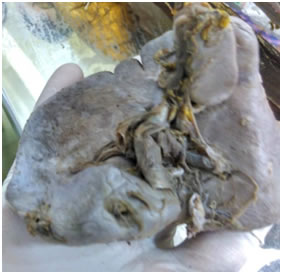
INTRODUCTION Spleen is a lymphoid organ situated in the upper left quadrant of the abdominal cavity between the fundus of stomach and diaphragm. [1] Spleen is mesodermal in origin. It begins to develop during fifth week of intrauterine life in the dorsal mesogastrium. Mesenchymal cells lying between the two layers of dorsal mesogastrium condense to form a number of small mesenchymal masses that later fuse to form a single mesenchymal mass.2 Any failure of this fusion results in small splenic tissues developing separately and resulting in accessory spleens. These accessory spleens are usually isolated but can be connected to the spleen by thin bands of similar tissue.3 Though most common site for accessory spleen is the hilum of spleen, they can be found in gastrosplenic and lienorenal ligaments also in pancreatic tail along the splenic artery. We found accessory spleen during routine dissection near the hilum of spleen. CASE-REPORT During routine anatomical dissection of abdomen in an adult male cadaver, we found an additional splenic mass near the hilum of spleen. This accessory spleen was connected with main splenic mass by a thin band of similar tissue. It was supplied by the branch of splenic artery and drained by a tributary from splenic vein. Main splenic mass was normal without any other variation. No other splenic tissue was found in gastrosplenic and lienorenal ligaments or in pancreatic tail.
Figure 1: Diagram showing accessory spleen
DISCUSSION The incidence of accessoryspleen, mentionedin the text book of Moore’s Anatomy is 10%.4 In India, Chaware PN5 reported 4.5% of the splenic samples withaccessory spleen while Mallikarjun6 in his cadaveric study found accessory spleen in 4 cadavers (8%) out of 50.In a study by Rayhan,7 the incidence of accessory spleen was 24.28% in Bangladeshi people while Romer T8 mentioned 10% of cases with accessory spleen in the study in Switzerland. Most accessory spleens are asymptomatic and incidental. They should not be confused with enlarged lymph nodes or masses in the adrenal gland or pancreas.9,10 Accessory spleens may result in interpretation errors in diagnostic imaging. In hematologic disorders, surgeons must be aware of accessory spleens so that they remove all functioning splenic tissue otherwise the hematologic disorders will recur.11 Hemorrhage and spontaneous rupture can also occur as a result of venous congestion and torsion of an accessory spleen.12,13 When an intra-abdominal mass is identified in a post splenectomy patient, even after lymph nodes dissection associated with gastric cancer, accessory spleen must be acknowledged as a differential diagnosis.14
REFERENCES
|
|
 This work is licensed under a Creative Commons Attribution-NonCommercial 4.0 International License.