 Home
Home|
Table of Content Volume 3 Issue 1 - July 2017
Study of essential parameters of foetal ovaries during intra uterine life
Teresa Rani S1, T Prasuna2*
1Associate Professor, Department of Anatomy, Guntur Medical College, Guntur, Andhra Pradesh, INDIA. 2Assistant Professor, Department of Anatomy, Kakatiya Medical College, Warangal, Telangana, INDIA. Email: thailamprasuna@gmail.com
Abstract Objective: To determine the development and the localization of the ovaries during the fetal period.Material and Methods: The material studied consisted of 42 normal human fetuses of different gestational age starting from 20 weeks to full term, collected from the following Hospitals of Warangal, Andhra Pradesh Chandrakantha memorial hospital, Warangal. Govt. maternity hospital, Hanumakonda. The following parameters are considered for the present study: The shape of ovaries, the positions of the ovaries, the localization of the ovaries with respect to linea terminalis, ureters, and the iliac arteries, the dimensions and the weight of the ovaries.Results: In the fetal period, the ovaries were most commonly almond shaped and had an oblique orientation. In the 2nd, 3rd trimester and full-term fetuses, it was observed that the ovaries were not in ovarian fossa, suggesting that decent of ovary was in progression during these times. During the intrauterine period, the ovaries were most commonly located anterior to the ureters and over the common iliac artery, towards the end of the 40th week, migrate to its final location between the internal and external iliac arteries. Conclusion: We found that the ovaries did not assume the position of the adults at the end of the fetal period, rather continued its descent after the birth. The right ovaries are larger than the left. We believe our findings about the fetal ovaries will be useful in obstetrics, fetal pathology, and forensic pathology.Key Words: Ovary, fetuses, positions, dimensions. Abbreviations: Right (Rt), Left (Lt), Crown rump length (CRL), Weeks (Wks), Fullterm (FT).
INTRODUCTION The ovary is a paired, female reproductive organ situated one on each side of the uterus close to the lateral pelvic wall and attached to the posterosuperior aspect of the broad ligament, and posteroinferior to the uterine tube by a double fold of peritoneum, the mesovarium1 The combined weight of the ovaries at birth is about 0.3gms. The ovaries are relatively large at birth and much larger than the testes2. In embryonic and early foetal life the ovaries are like the testes, situated in the lumbar region near the kidneys, but they gradually descend into the lesser pelvis2 from 20 weeks to 28 weeks, the ovaries were located in the false pelvis on either side of the sigmoid colon. Anteriorly they were related to the corresponding uterine tube and posteriorly they were related to the ureter and common iliac artery. At 40th week both ovaries were placed obliquely on each side of the terminal part of sigmoid colon. Laterally there were related to the corresponding uterine tube. Medially both the ovaries were in contact with the sigmoid colon.
MATERIAL AND METHODS The following are the material utilized for the present study. Forty two dead fetuses of different age groups have been collected from the following Hospitals of Warangal, Andhra Pradesh Chandrakantha memorial hospital, Warangal. Govt. maternity hospital, Hanumakonda. All the specimens were either aborted, still born or premature deliveries resulting in neonatal death. All the fetuses were preserved in 10% formalin after injecting 20cc to 100cc of 10% formalin into cranial cavity, thorax and abdomen depending on the size of the fetus. After embalming, the fetuses were immersed in buckets containing 10% formalin. Dissection of fetuses was taken up 48 hrs after embalming. This is to ensure that all the structures are fixed in situ. The following parameters have been included as they are essential for future analysis. Length all fetuses has been measured in cm. i.e., the crown rump length to calculate approximate age of fetuses in weeks. The weight of fetus along with 5 cm – 6 cm of umbilical cord have been taken in grams with the help of simple balance. Biparietal diameters i.e., distance between two parietal eminences of all specimens were measured with vernier calipers. The age of the fetuses were calculated from the crown rump length (CRL) and gross features. To expose the ovary, a systematic dissection procedure has been adopted. The ovaries were removed by cutting the ligaments and were observed in detail. OBSERVATION AND RESULTS Both ovaries were seen in all age group in the false pelvis on naked eye examination as whitish elongated almond shaped structure on either side of the sigmoid colon. The cranial portion of the ovary was found to be related to the ampulla of the uterine tube and caudally it pointed to the junction between uterus and uterine tube. Its posterior relations were ureter and common iliac artery. Parameters of both right and left ovaries were obtained from 42 fetuses of different age groups ranging from 20 weeks to full term. Mean of all parameters i.e., length, width, thickness and weight were calculated for each ovary and shape of both right and left ovaries were noted and compared with that of available previous studies. For the convenience, the collected fetuses were divided in to four groups. Group – I 20-24 weeks, Group-II 25-28 weeks, Group-III 29-32 weeks, Group-IV 33 weeks to full term.Table 1
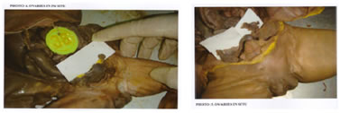
Figure 1 Figure 2 Figure 3 Figure 4 Figure 1: 24 wks fetus with ovaries in situ; Figure 2: 28 wks fetus with ovaries in situ; Figure 3: 32 wks fetus with ovaries in situ; Figure 4: 38 wks fetus with ovaries in situ
DISCUSSION In the neonate the ovaries are found in the lower part of the iliac fossae. They complete their descent into the ovarian fossae in early childhood. The long axis of the ovary is almost vertical in the neonate, becoming temporarily horizontal during descent and vertical once more in the ovarian fossa. V. Usha Rani et al reported in their study that of 80 ovaries all prenatal ovaries were located at the pelvic brim except 2 smallest embryo in which they were in lumbar region3. Osman sulak et al reported trimesterwise variations in the location of the fetal ovaries starting from the pelvic brim indicating a gradual descent with progress in gestational age4. In the prenatal group, the ovaries were placed transversly and in postnatal group in oblique or horizontal position. Variability in position has been reported to be due to the state of surrounding organs5. In the prenatal group, the predominant shape was oval in all 3 gestational age groups studied3. Osman sulak et al reported predominance of almond shape in Turkish fetuses4. There are no reports on percentage incidence of various ovarian shapes available in literature although almond and oval shapes have been reported4,6. Prenatal and postnatal ovaries were dull white, pink yellowish or gray in color7.The observations on color of ovaries in this study are in agreement with the earlier report on this particular morphological feature7. The observations in this study on length, width, and thickness of ovaries are comparable with earlier reports (table-2 ). The length of ovaries during 20-40 weeks in this study are in agreement with that reported by osman sulak et al4. Similarly, the increase in width and thickness of the ovaries during this period in study is in agreement with earlier reports4. The combined weight of ovaries at birth is about 0.3gms2. Our observations of weight of ovaries are in agreement with that reported in the literature. As per the study of Ann Hum Genet who did a study on differential growth of human fetal gonads with respect to sex and body side, has observed that the mean values of gonads are more on right side than on the left8. According to Mitt Who et al, in comparative measurement of weight of human fetal gonads investigated values found that right ovary values are greater than left ovary9. In the present study the mean weight of ovary (grams) was as follows: Table 2
The above table show that the mean values of right ovary weights are more than the left ones. It is in correlation with the statement of the first two authors.
Table 3
The present study shows that length, width and thickness of ovaries were found to be increasing with fetal gestational age. The mean length of Rt and Lt ovary in fetuses of 20 weeks to Full term was 1.0 cm, the mean width of Rt and Lt ovaries in fetuses of 20 wks to Full term was 0.79 cm, the mean thickness of right and left ovaries in fetuses of 20 weeks to Full term was 0.35 cm and the mean weight of right and left ovaries in fetuses of 20 week to Full term was 0.37 grams which are very close to available literature.
CONCLUSION Prenatal ovaries were found to be oval or almond shaped and were present in false pelvis before birth. The detailed knowledge of developmental anatomy of ovaries will be useful in obstetrics, fetal pathology, and forensic pathology.
REFERENCES
|
|
||||||||||||||||||||||||||||||||||||||||||||||||||||||||||||||||||||||||||||||||||||||||||||||||||||||||||||||||||

 This work is licensed under a Creative Commons Attribution-NonCommercial 4.0 International License.