 Home
Home|
Table of Content Volume 3 Issue 1 - July 2017
Evaluation of degenerative changes in lumbar spine by MRI in rural population
Walwante R D1, Dhapate S S2*, Porwal S S3
1,3Assistant Professor, 2Professor and HOD, Department of Anatomy, S.R.T.R.G.M.C. Ambajogai-431517, Dist. Beed, Maharashtra, INDIA. Email: walwanteraviraj@gmail.com, dhapatess@rediffmail.com, satish27181@yahoo.coo.in
Abstract Background and Aims: Degeneration of inter vertebral disc is characterized by a loss of hydration of the nucleus pulposus and disruption of the annulus fibrosus resulting from a variety of mechanical and biochemical factors. Individuals with degenerative disease of the lumbar spine can be symptomatic or asymptomatic, although commonly the disease is asymptomatic. The symptomatic individuals can present with back pain or radicular pain syndrome. Lumbar spine is the common area affected by degenerative changes, as it is a part of spine which is subjected to heavy mechanical stress. Material and Methods: This is a hospital based cross-sectional descriptive study. Study population included all patients above 20 years of age with LBP with/without radiculopathy who were referred for lumbar spine MRI at Radiology department.. Observations and Results: The study included 165 patients; the age range was from 20 to 80 years whereby 49.69% of them were females. On lumbar MRI, overall prevalence of lumbar degenerative findings was disc bulge 76%, nerve root compression 40%, and central canal stenosis 26%. The least common finding was disc herniation which was seen in 18%. 9% participants had normal lumbar MRI findings. Prevalence of various degenerative imaging findings was more common among males, only disc bulges were common among females, though the differences were not statistically significant. Conclusion: Degenerative changes in lumbar spine increase with age, being more common among males than females and there is strong association between low back pain patients and degenerative changes observed on MRI. Key Words: Back pain, disc degeneration, disc displacement.
INTRODUCTION Ageing is main factor implicated in spine degenerative disease. Its prevalence increases with age. It ranges from 85% to 95% among adults aged 50 to 55 years, with no sex difference 1, 2.Hult estimates that up to 80% of population is affected by this symptom at some time in life3.The symptomatic individuals can present with back pain or radicular pain syndrome4. Individuals with degenerative disease of the lumbar spine can be symptomatic or asymptomatic, although commonly the disease is asymptomatic5-8. Back pain is strongly associated with degeneration of the intervertebral disc7. The symptomatic individuals can present with back pain or radicular pain syndrome (sciatica) 9. The possible sources of pain are mechanical compression of neural elements by disc herniation, as well as direct biochemical and inflammatory 6,9. Asymptomatic individuals may have degenerative spine findings, including: disc degeneration, disc bulges, and spinal stenosis5,7,8. Apart from age other factors have been implicated as causes of spine degenerative disease, these include; genetic inheritance, physical loading history, trauma and impaired nutrition4,10. Lumbar spine is the common area affected by degenerative changes, as it is a part of spine which is subjected to heavy mechanical stress11. Despite the ambiguous relationship between anatomy and pain, clinicians rely heavily on medical imaging to identify patho-anatomic changes. One of the most important advances relating to the evaluation of the intervertebral disc (IVD) comes from the high-resolution in vivo images of the lumbar spine obtained by magnetic resonance imaging (MRI). Recent MRI studies of people with or without LBP have yielded surprising results. The role of diagnostic imaging in spine degenerative disease is to evaluate the status of the neural tissues and to affect the therapeutic decision making12.Imaging is only justified in patients for whom surgery is considered. The commonly used imaging modalities are plain film, CT and MRI. Plain film examination of the lumbar spine is the usual initial imaging technique13.Plain radiography provides only limited diagnostic information because it cannot show the structural morphology of the intervertebral disc.MRI has become the primary imaging modality for evaluation of degenerative disorders of lumbar spine. The intervertebral disc, vertebrae, ligaments, spinal canal and neural foramina may also be best evaluated using current MRI techniques. In this study the anatomical cross-sectional imaging and disco-graphic findings of patients who are referred for MRI for low back pain have been reviewed.
MATERIAL AND METHODS This is a hospital based cross-sectional descriptive study. Study was done for estimating pattern of degenerative changes seen in patients with low back pain referred to lumbar magnetic resonance imaging centre located in rural area. A total of 175 individuals had lumbar MRI scan in year, but only 165 whom fulfilled the study criterion were studied. Those patients above 20 years of age, with history of low back pain with/without radiculopathy are included in this study. Those patients having history of former lumbar spine surgery, vertebral trauma, and spine tumour and spine infection were excluded form study. Permission to conduct the study at Radiology department was obtained from authority. Imaging was performed by a trained Radiographer. Lumbar spine MRI was done using 0.03OTscanner (Xin Ao MDT). The findings assessed on MR imaging were Disc bulge, Disc herniation, Central canal stenosis and Nerve root compression. The clinical condition of the subjects collected from data was compared with the imaging findings. Data analysis was done by using the software statistical package for social sciences (SPSS) software and using Open Source epidemiological statistics for public health version 2.3.1.
OBSERVATIONS AND RESULTS The study included 165 patients, the age range was from 20 to 80 years (mean; 47.49 14.33 years) whereby 82 of them were females and 83 were males. (Table 1).
Table 1: Shows age group and sex of patients included in study
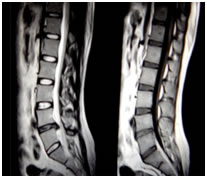
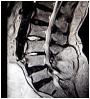
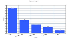
On lumbar MRI, overall prevalence of lumbar degenerative findings was disc bulge (figure 2) 125 (75.75%), nerve root compression 66 (40%), and central canal stenosis 44 (26.66%). The least common finding was disc herniation (figure 1) which was seen in 31 patients (18.78%) (Fig.1and2). Minority of participants 15 (9.09%) had normal lumbar MRI findings (Fig. 3). Figure 1: Shows degeneration of intervertebral disc at L4/L5 level with disc herniation and compression of cord
Figure 2: Shows degeneration of lumbar intervertebral discs and at L4/L5disc herniation with sequestration and cord compression. Disc budges at L5/S1 level Figure 3: Graphical presentation of degenerative findings The prevalence of lumbar degenerative findings was increasing with age. This was true for disc bulge, central canal stenosis, nerve root compression and (P-values; 0.000, 0.018 and 0.013 respectively) for disc herniation the prevalence varied with age but the differences were not statistically significant (p- value > 0.05). (Table 2).
Table 2: Degenerative Findings In Different Age Group
Prevalence Of Various Degenerative Imaging Findings Was More Common Among Males but disc bulge were common among females, though the differences were not statistically significant (p-value ≥0.05).(Table 3).
Table 3: degenerative findings in different sex
Most of the degenerative findings were seen at lower lumbar levels i.e. L4/L5 and L5/S1. At L4/L5 the prevalence of disc bulge, disc herniation, central canal stenosis and nerve root compression were 117(70.90%), 20(12.12%), 31(18.78%) and 41(24.84%) respectively, whereby these findings at L1/L2 were; 3(1.81%), 0(0%), 0(0%) and 0(0%) respectively. (Table 4). Table 4: Degenerative findings at different disc level
(DB=DISC BULGE, DH= DISC HERNIATION, CS= CANAL STENOSIS, NRC= NERVE ROOT COMPRESSION) Percentage distribution of type of disc herniation (n=40: total number of herniated discs, (percentage in brackets) 27(67.5%) of all herniated discs were extrusions. Only 13 (32.5%) discs were protrusions.(Table 5).
Table 5: Shows type of disc herniation
Percentage distribution of the location of herniated. (n=40: total number of herniated discs)(Percentage in bracket) (Table 6). Table 6: Shows location of disc herniation
The most common location for disc herniation was postero-lateral seen in 30 (75%) discs, followed by posterocentral and foraminal 7 (17.5%) and 3 (7.5%) respectively, so the intraspinal disc herniation (post central and posterolateral) were the most common, seen in 37 (92.5%) herniated discs. Prevalence of disc bulge did not significantly vary with patient presenting symptoms. Disc herniations, central canal stenosis and nerve root compression were common in patients with radiculopathy than in patients with LBP only. The prevalence was 24 (32.43%), 42 (56.75%), 60 (81.08%) respectively for patient with radiculopathy and 7 (7.69%), 2 (2.19%), 31 (34.06%) respectively for patient with LBP only (p-value: 0.000). only 2(2.19%) patients with low back pain only had canal stenosis. (Table 7).
Table 7: Degenerative findings in symptomatic patients
(LBP= Low back pain) DISCUSSION The role of diagnostic imaging is to provide accurate anatomical information and to affect the management decision making. This cross-sectional hospital based study used MRI to diagnose spine degenerative changes as it has better tissue segregation and it can show degenerative changes at an early stage as compared to other imaging techniques (such as CT scan)12. Other advantage of MRI includes having no known side effects or morbidity, no radiation exposure and is non-invasive12,14,15. Despite its high sensitivity, degenerative changes are observed on many MRI scans in asymptomatic subjects, thus questioning its specificity14. That’s why MRI is only beneficial to patients with chronic disease and those who are being planned for spine surgery. All recruited patients underwent MRI of the lumbar spine and both sagittal and axial views of all images were interpreted to locate the degenerative findings. Degenerative changes were observed in majority of patients examined. Most of these degenerative findings were seen at L4/L5 and L5/S1. Though a degenerative change of the disc begins early in life and is partly a consequence of aging, the actual cause is not known but many factors (autoimmune, genetic, re-absorption and biochemical) have been implicated in accelerating the process. Since lumbar spine is subjected to heavy mechanical stress, it is a common area affected by degenerative changes16; this could partly explain such observation in this study group. The mean age of this study group is 47.49 14.33 years, could be another explanation, as degenerative changes is common in individuals above 40 years of age and its prevalence increases progressively to over 90% by 50 to 55 years of age17-19,. Disc bulge was the most frequent finding observed in 125(75.75%) patients in this study. The difference observed between the age groups was significant (p-value= 0.005) and compares with the findings of other previous studies 2, 17-19. The prevalence of disc degeneration to young individuals (20 to 39 years) could probably be explained as results of genetic predisposition; though, other factors like repeated traumatic injuries and physical loading history can play a role in causing disc degeneration2,8,17,19. The difference in prevalence among young and aged individual could be contributed by aging process. These radiological findings should receive more emphasis during the interpretation of MR images of patients who present with radiculopathy. Though In this study MRI has revealed high frequency of lumbar degenerative imaging findings; MRI may also reveal high rates of abnormalities in asymptomatic individuals14. About 35% of asymptomatic individuals have abnormal MRI findings5,7,8,20. Because of these findings among asymptomatic individuals, MRI alone cannot be used to define the cause of symptoms among symptomatic patients. MRI findings must be correlated with patient age, clinical signs and symptoms following a careful physical examination for accurate management decisions.
CONCLUSION There is a strong association between low back pain patients and degenerative changes in lumbar spine observed on MRI. Degenerative changes in lumbar spine increase with age, being more common among males than females. 90% of studied patients show degenerative findings on MRI. Disc budge is the most frequent finding. The least finding is disc herniation, whereby extrusion is more common and it is commonly located posterolateral. The patients who were having LBP with radiculopathy shows disc herniations, canal stenosis and nerve root compression and the patients who presented with LBP only shows more commonly disc bulge finding.
REFERENCES
|
|
||||||||||||||||||||||||||||||||||||||||||||||||||||||||||||||||||||||||||||||||||||||||||||||||||||||||||||||||||||||||||||||||||||||||||||||||||||||||||||||||||||||||||||||||||||||||||||||||||||||||||||||||||||||||||||
 This work is licensed under a Creative Commons Attribution-NonCommercial 4.0 International License.