|
Table of Content Volume 3 Issue 2 - August 2017
Morphometric analysis of human sacra
Anant Karbhari Shingare1*, N B Masaram2, S S Dhapate3
1Assistant Professor, 3Professor and HOD, Department of Anatomy, S.R.T.R. Government Medical College, Ambajogai, Maharashtra, INDIA. 2Assistant Professor, Department of Anatomy, Dr Vaishampayan Memorial Government Medical College, Solapur, Maharashtra, INDIA. Email: anant.karbhari@gmail.com, masaramnitin9@gmail.com
Abstract Background: Sacrum is formed by fusion of five sacral vertebrae and forms the caudal end of the vertebral column. Anatomists and anthropologists since long acknowledged the importance of sacrum in identifying the sex of a deceased person. Sexual dimorphic characters can be studied both morphologically and metrically. Material and Methods: The present study was performed at Department of Anatomy, S. R. T. R. Government Medical College, Ambajogai, Maharashtra on 50 (25 male and 25 female) adult human sacra of known sex. Following equipments used were Sliding verniercaliper, divider, steel Measuring Scale. Parameters studied were Maximum length of sacrum, Maximum breadth of sacrum, Sacral index. Aims and Objective: This study was conducted to determine sexual dimorphism of adult sacrum, to evaluate the most significant parameter in sexual dimorphism and also to compare and contrast the result of present study with previous studies. Results and Conclusion: Ventral straght length and sacral index was found to be highly significant with a p value of <0.0001.Maximum breadth was found to be not significant with a p value of <0.0566.From the present study we find out similarities and differences in the metrical values of different sacral parameters in males and females and also highlighted the best parameter which can be used for sexual dimorphism of sacrum. Key Words: Morphometric, human sacra.
INTRODUCTION Sacrum is formed by fusion of five sacral vertebrae and forms the caudal end of the vertebral column. The sacrum supports the erect spine, provides the strength and stability to the bony pelvis in transmitting body weight. The bones of body are the last to perish after death next only to the enamel of teeth. Determination of sex is an integral first step in the development of the biological profile in human osteology. Sex determination is necessary to make age, ancestry and stature estimations, as the sex's age differently, exhibit some degree of variation in ancestry related morphology and generally differ in height. (Kothapalli). Anatomists and anthropologists since long acknowledged the importance of sacrum in identifying the sex of a deceased person.
MATERIALS AND METHODS The present study was performed on 50 (25 male and 25 female) adult human sacra of known sex. All of them were dry and free from deformity and fully ossified. All the sacra were obtained from Department of Anatomy, S.R.T.R. Government Medical College, Ambajogai, Maharashtra Following equipments were used for measurement of various parameters.
Each sacrum was studied for different features of sexual dimorphism and sacral hiatus. Sexual dimorphism
Sacral index = (sacral width / sacral ventral straight length) x 100. After completing measurements, they were tabulated and statistically analysed with the help of SPSS software. Mean, standard deviation, range, demarking points and percentage of identified bones calculated.
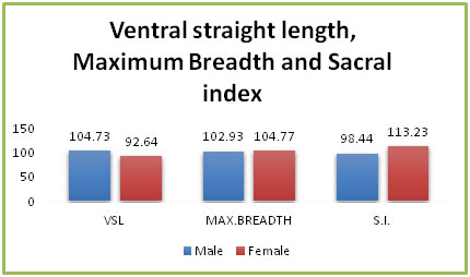
RESULTS At the first of study, each parameter were tabulated and statistically analyzed. Comparative graphs of male and female values were drawn which shows the zone of difference and overlapping between male and female values. The ventral straight length: The ventral straight length was found to be having a mean of 104.73 in males and 92.64 in females. The demarking points for males was >110.94 and <86.91 in females. This parameter was helpful in identification of 16.86% of bones in males and 19.30% of bones in females. Ventralstraght length was found to be highly significant with a p value of <0.0001 The maximum breadth: The maximum breadth was found to be having a mean of 102.93 mm in males and 104.77 mm in females. The demarking points for males was >124.21 and <88.44 in females. Maximum breadth was found to be not significant with a p value of <0.0566. Sacral Index: The sacral index was found to be having a mean of 98.44 mm in males and 113.23 mm in females. The demarking points for males was <96.4 and >112.51 in females. This parameter was helpful in identification of 27.71% of bones in males and 57.9% of bones in females. Sacral index was found to be highly significant with a p value of <0.0001. Table 1: Showing measurements of Ventral straight length, Maximum breadthand sacral index in males and females
Figure 1: Showing measurements of all three parameters DISCUSSION Table 2: Ventral Straight Length
N= Sample size, X= Mean, S.D.= Standard deviation, p= Probability, N.S.= Not significant, S.S.D= Statistically significant difference between two sexes
Table 3: Maximum breadth
Table 4: Sacral Index
Various studies of different authors were compared with present study for all three parameters are shown in Table 2,3 and 4. CONCLUSION The mean value for ventral straight length was found to be104.73 mm in males and 92.64 mm in females and was statistically highly significant. The study is correlated with Raju et al and Sachdeva et al. The mean value for maximum breadth found to be 102.93 mm in males and 104.77 mm in females and was statistically not significant. 0% of bones were identified by using this parameter. The findings of present study correlated with study done by Raju et al and Sachdeva et al. The mean value for sacral index found to be 98.44 mm in males and 113.23 mm in females. 57.9% of bones in females and 27.71% of bones in males were identified by using this parameter. This is most significant parameter for sex determination. Similar findings were reported by other research workers such as Mishra et al, Shailaja et al, Patel. Anatomists and anthropologists since long acknowledged the importance of sacrum in identifying the sex of a deceased person. The present study was undertaken to find out similarities and differences in the metrical values of different sacral parameters in males and females and also highlight the best parameter which can be used for sexual dimorphism of sacrum.
REFERENCES
|
|
||||||||||||||||||||||||||||||||||||||||||||||||||||||||||||||||||||||||||||||||||||||||||||||||||||||||||||||||||||||||||||||||||||||||||||||||||||||||||||||||||||||||||||||||||||||||||||||||||||||||||||||||||||||||||||||||||||||||||||||||||||||||||||||||||||||||||||||||||||||||||||||||||||||||||||||||||||||||||||||||||
 Home
Home

