 Home
Home|
Table of Content Volume 10 Issue 1 - April 2019
A cadaveric study of the third head of biceps brachii in south Indian population
P R Prefulla1, B J Bhuvaneswari2*
1,2Assistant Professor, Institute of Anatomy, Madras Medical College, Chennai, PIN- 600003, INDIA. Email: prefullanambiar@gmail.com, bhuvanaranjith04@gmail.com
Abstract Biceps brachii is a two headed muscle in the flexor compartment of arm. Third head of Biceps brachii has been reported by various authors worldwide. The third head of Biceps brachii has significant functional and clinical implications. The present study was carried out in 100 upper limb specimens from 50 embalmed adult human cadavers at the Institute of Anatomy, Madras Medical College, Chennai, India. Presence of third head of Biceps brachii, its origin, insertion and nerve supply were studied in detail. The findings would be valuable to Anatomists, Radiologists, Orthopaedicians and Plastic surgeons. Key Word: Biceps brachii, Third head, Musculocutaneous nerve, Intramuscular compression, Biceps brachii flap, Fracture humerus.
INTRODUCTION Biceps brachii is a large fusiform muscle in the flexor compartment of arm. The muscle arises by two heads, as implied by its name. The tendon of long head is attached to the supraglenoid tubercle of scapula and lies intracapsular in the shoulder joint. The short head arises as a thick flattened tendon from the tip of coracoid process of scapula. The tendinous origin of each head expands into fleshy bellies. The two bellies lie side by side, loosely connected by areolar tissue, but do not merge to form a single flattened tendon until just above the elbow joint. The tendon of Biceps brachii gets inserted to the posterior part of radial tuberosity8. Bicipital aponeurosis spreads downwards and medially from the anteromedial aspect of the tendon and partly in continuity with the fleshy fibres of the muscle. It joins the deep fascia of forearm and is inserted into the subcutaneous border of the upper end of ulna11. Each head of Biceps brachii receives a twig from Musculocutaneous nerve. These twigs are composed of fibres derived from the ventral rami of 5th and 6th cervical spinal nerves19. Biceps brachii is a three-joint muscle- crossing the shoulder, elbow and superior radio-ulnar joints11. It is a powerful supinator. It flexes the elbow, most effectively with forearm supinated and also acts as a weak flexor of the shoulder joint8. A not uncommon variation of the Biceps brachii muscle is the presence of a third head19. The origin of third head of Biceps brachii is variable. It may be from the shaft of humerus near the insertion of coracobrachialis6, or fascia over brachialis7, intertubercular sulcus3, anterior limb of V shaped insertion of deltoid5. The presence of a third or humeral head might increase the dynamics of Biceps brachii, as otherwise the muscle is devoid of any humeral attachment. This may cause unusual bone segment displacement in fracture of humerus. The incidence of third head of Biceps Brachii has been reported by several authors worldwide. The present study was carried out to explore the third head of Biceps brachii in South Indian population.
MATERIALS AND METHODS The study was carried out in 100 upper limb specimens from 50 embalmed adult human cadavers allotted for routine undergraduate dissection at the Institute of Anatomy, Madras Medical College, Chennai, India. Careful dissection was done to study the Biceps brachii muscle from proximal to distal attachments. Presence of third head of Biceps brachii, its origin, insertion and nerve supply were analysed in detail.
OBSERVATIONS AND RESULTS Out of 100 upper limb specimens, 11 showed the presence of third head of Biceps brachii (Fig.1). It was noted bilaterally in one cadaver. Out of the remaining 49 cadavers, third head was noted on the right side in 5 upper limbs and left side in 4 upper limbs. Table 1: Cadaveric incidence of third head of Biceps brachii
Table 2: Site of origin of third head of Biceps brachii
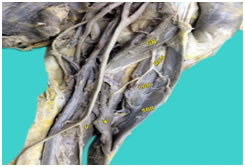
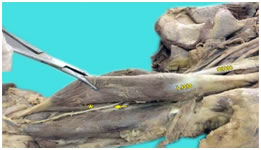
In all cases, the third head of Biceps brachii merged with the other two heads (Fig.1) and inserted through the tendon of biceps brachii and bicipital aponeurosis. The third head was innervated by a twig from Musculocutaneous nerve. Presence of third head was not associated with any variation in the long and short heads of Biceps brachii. In 3 upper limbs (3%), Musculocutaneous nerve was found to course between third head and the other 2 heads. (Fig.2) Figure 1: Left front of arm showing Long head of Biceps brachii (LBB), Short head of Biceps brachii (SBB) and Third head of Biceps brachii (*). Note the origin of Third head of Biceps brachii from shaft of humerus, from the area between insertion of Coracobrachialis (CB) and origin of Brachialis (B). Note the nerve supply to all the three heads of Biceps brachii from Musculocutaneous nerve (MCN). Figure 2: Left front of arm showing Musculocutaneous nerve between Third head of Biceps brachii (*) and the other two heads. LBB- Long head of Biceps brachii, SBB- Short head of Biceps brachii.
DISCUSSION In the present study, the incidence of third head of Biceps brachii was 11%. This correlates to the reports of Amar Jayanthi et al(10.8%)2 and Ramakrishna Avadhani et al (16.67%)12 In 10 out of the 11 upper limbs in which third head of Biceps brachii was present, it took origin from shaft of humerus. The site of origin was the area between insertion of Coracobrachialis and origin of Brachialis, coinciding with the findings of Prabhjot Cheema et al10, Abdullah G1, PP Poudel et al9, Renu Gupta et al14 and Renata Pacholczak et al13. Origin from fascia over Brachialis, which was 1% in the present study, has also been reported by Vinnakota Sunitha et al18 and S. Soni et al16. The other reported areas of origin of the third head are intertubercular groove of humerus3 and anterior limb of V shaped insertion of Deltoid5. These were not found in the present study. In the present study, unilateral incidence of third head of Biceps brachii(10%) was found to be much higher than bilateral incidence(1%). This is similar to the findings of Amar Jayanthi et al2, Prabhjot Cheema et al10 and Eti Sthapak et al4. The incidence was found to be slightly higher on the right side (6%) than on the left side (5%). This is similar to the findings of Eti Sthapak et al4. In all cases, the third head of Biceps brachii joined with the other 2 heads and inserted through Biceps tendon and bicipital aponeurosis. This harmonizes with the findings of B.O Donmez et al3, Satheesha Nayak et al15 and Eti Sthapak et al4. Nerve supply of the third head was by a twig from Musculocutaneous nerve, in all cases. This conforms to Prabhjot Cheema et al10, Vinnakota Sunitha et al18, Hitendra Kumar5 et al and B.O Donmez et al3. Musculocutaneous nerve was found to course between the third head and the other two heads in 3%. This tallies with the findings of M. Mujahid Ansari et al7 and Satheesha Nayak et al15. This intramuscular course is a potential site for compression of musculocutaneous nerve.
CONCLUSION Knowledge of the incidence of third head of Biceps brachii and its morphology is of importance in traumatology. The intramuscular course of Musculocutaneous nerve gains importance in hypertrophied Biceps brachii muscle as in professional body builders and weight lifters. Biceps brachii muscle flaps are used in plastic and reconstructive surgeries for axillary wound coverage. An additional head would be of added value in such cases. Hence the findings of the present study would be valuable to Anatomists, Radiologists, Orthopaedicians and Plastic surgeons.
REFERENCES
|
|
|||||||||||||
 This work is licensed under a Creative Commons Attribution-NonCommercial 4.0 International License.