 Home
Home|
Table of Content Volume 10 Issue 1 - April 2019
Determination of sex from greater sciatic notch of hip bone: A cross sectional study at tertiary care hospital in Maharashtra
K Thomas Manoj1, RD Walwante2*, MM Baig3
1,2Assistant Professor, Department of Anatomy, SRTR Government Medical College, Ambajogai, Maharashtra, INDIA. 3 Professor & Head, Department of Anatomy, Government Medical College, Latur, Maharashtra, INDIA. Email: telmaha2710@gmail.com
Abstract Background: Identification of sex of an unknown individual from the skeletal remains is the first and the most important step encountered by the forensic experts and archeologists. The hip bone is considered as an ideal bone for sex determination as it provides the highest accuracy levels. Objectives: The present study is done to implicate the importance of greater sciatic notch in sexing skeletal remains from Maharashtra region of India by morphometric study. Material and Methods: One hundred and twenty adult hip bones of known sex including 70 male and 50 female hip bones were numbered and used for the study. All these were obtained from skeletal collection of Department of Anatomy across all medical colleges in Maharashtra. Care was taken to avoid damaged and pathologically deformed bones that could lead to error in measurement. Results: There was statistically significant difference in the Width, Depth, Anterior Segment Posterior Segment, Three Indices, Posterior Angle, Total Angle of male and female greater sciatic notch, whereas Anterior angle of greater sciatic notch is statistically insignificant. Conclusions: Present study not only gives understanding of the bisexual dimorphism but also is much applicable in medico legal aspect and archeology. Key Word: Greater sciatic notch, Sexing of hip bone.
INTRODUCTION Determination of sex of an unknown individual is one of the critical questions addressed when human skeletal remains are found both in forensic investigation and studies of past population. Therefore the study of sexual dimorphism of bones in human population is a matter of interest not only for Anatomists but also for the Anthropologists and Forensic experts1.The nature has allowed the individual anatomical variation and departures from the set norms within each sex. In addition, these variations are affected by multiple etiological factors such as cultural, environmental and genetic elements 2 Hip bone is an ideal bone for sex determination because it not only reflects the general differences between the two sexes but also the special adaptation of female hip bone for child bearing. The greater sciatic notch is especially valuable in such situations because it is highly sexually dimorphic and resistant to damage thus can often be scored in poorly preserved skeletons. The introduction of metric method or precise measurement method has provided the simplicity and accuracy to determine the sex of skeletal remains. Techniques which require the measurement of diameters, circumferences or cross sectional areas of tubular bones may provide the needed means for sexing fragmentary remains4. Various studies have resulted a wide range of variation in morphological and morphometric sex differences because the skeletal growth is influenced by factors like race, heredity, climate, nutrition and environment which changes from region to region. Hence there is a need to study it different population. The present study is done to implicate the importance of greater sciatic notch in sexing skeletal remains from Maharashtra region by morphometric study.
MATERIAL AND METHODS One hundred and twenty adult hip bones of known sex including 70 male and 50 female hip bones were numbered and used for the study. All these were obtained from skeletal collection of Department of Anatomy across all medical colleges in Maharashtra. Care was taken to avoid damaged and pathologically deformed bones that could lead to error in measurement. The following adult human hip bones which are included for the study are fully ossified, not broken, not having any deformities with intact Greater sciatic notch, ischial spine, and Posterior inferior iliac spine. The sampling of data was made randomly. The materials used for this study includes digital vernier calipers protractors. In present study following measurements were taken on these hipbones. The definitions of the landmarks and measurements were taken from the literature and were selected on the basis of their being good discriminators in previous studies. Each variable was measured three times and the mean value of three measurements was calculated for each variable for each hipbone.
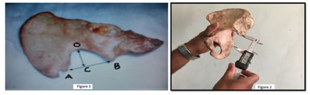
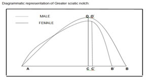
Measurements of greater sciatic notch Figure 1: The point A was taken as Posterior inferior iliac spine and point B was taken as ischial spine; Figure 2: Maximum width (AB) of greater sciatic notch
It is the maximum distance between the posterior inferior iliac spine and the tip of ischial spine Figure 3: The curvature of greater sciatic notch was then plotted on a paper. From the deepest point (O) of sciatic notch perpendicular line was drawn to the base line which meets at (C). Then (OC) was taken as the depth of sciatic notch. Then (BC) was taken as Anterior segment (AS) and (AC) was taken as Posterior segment. Anterior angle BOC, Posterior angle AOC and Total angle AOB were measured with protractor after construction of a triangle on a paper from the above. Following formulae were used to calculate various indexes. INDEX I = ×100 INDEX II = ×100 INDEX III = ×100
Figure 4: Diagram shows various angle used in this study. RESULTS Table 1: Statistical analysis of width (AB) and depth (BC) of greater sciatic notch
There was statistically significant difference between the width and depth of male and female greater sciatic notch.
Table 2: Statistical analysis of anterior (BC) and posterior (AC) segment of of greater sciatic notch
Both anterior and posterior segment of GSN is statistically highly significant.
Table 3: Statistical analysis of various indexes of greater sciatic notch.
All the indexes were highly significant.
Table 4: Statistical analysis of various angles of greater sciatic notch
Anterior angle of GSN is statistically insignificant, rest posterior and total angles were highly significant DISCUSSION As a general rule male bones are more massive and heavier than female bones. The crests, ridges, tuberosities and lines of attachment of muscles and ligaments are more strongly marked in males. This rule also governs the size of joints and articular surfaces as well. It is generally recognized that of all the elements of human skeleton the in nominate bone offers best prospect for identification of sex. Unfortunately, the features of in nominate that exhibit highest levels of sexual dimorphism are frequently found to be damaged or missing in exhumed material According to Davivongs V et al5, Singh et al6 the width of greater sciatic notch is greater in females than in males. So also in the present study. According to Davivongs V et al5 the depth of greater sciatic notch is greater in females than in males. But a study done by Jovanovic S et al7, Singh et al6 the depth of greater sciatic notch is greater in males than in females. Thus, in the present study also there is a difference between the means which are statistically highly significant. The mean value of anterior segment of greater sciatic notch was found to be higher in females proving it to be statistically highly significant. According to Davivongs V et al5, Singh et al6the length of posterior segment of greater sciatic notch is greater in females than in males. The present study also shows the same results proving it to be statistically highly significant which can be used for sexing of hipbone. According to Singh et al6, Shah S et al8 and Suma et al9 the index I (depth (OC) / width (AB) ×100) is higher in males than in females and the index II (posterior segment (AC) / width (AB) ×100) is higher in females than in males. The present study also shows the same results. Thus the difference between two is statistically significant and this parameter has a high use for sexing of hipbone. According to the present study the mean value of index III (anterior segment (BC) / width (AB) ×100) was found to be higher in females. The difference between the two is statistically significant. Thus this parameter is of a little value in sexing of hipbone. According to the present study there is hardly any difference in the means of anterior angle rBOC of Greater sciatic notch of both males and females. Hence the means of males and females are statistically insignificant. Thus this parameter has no value for sexing of hipbone. According to Singh et al6, Takahashi H et al10, Rajarshree et al11, Shah S et al8and Suma et al9the posterior angle of greater sciatic notch r AOC is higher in females than in males. The present study also shows the same results. The difference between two is statistically significant. Thus this parameter has a high use for sexing of hipbone. According to Washburn SL et al12, Singh et al7, Takahashi H et al10, Rajarshree et al11 , Shah S et al8and Suma et al9the total angle of greater sciatic notch r AOB is higher in females than in males . The present study also shows the same results. The difference between two is statistically significant and this parameter has a high use for sexing of hipbone.
CONCLUSION Width, Depth, Anterior segment, Posterior segment, three indexes, posterior and total angle of greater sciatic notch was associated with a significant change in sexing the hip bone. Anterior angle was poor sex indicator which could not determine the sex of hip bone. Thus the present study not only gives understanding of the bisexual dimorphism but also is much applicable in medico legal aspect and archaeology.
REFERENCES
|
|
|||||||||||||||||||||||||||||||||||||||||||||||||||||||||||||||||||||||||||||||||||||||||||||||||||||||||||||||||||||||||||||||||||||||||||||||||||||||||||||||||||||

 This work is licensed under a Creative Commons Attribution-NonCommercial 4.0 International License.