 Home
Home
|
Table of Content - Volume 19 Issue 1 - July 2021
An osteological study of distal end of femur in adult south Indian population
Ashwini N S1, Asharani S K2, Jyothi Lakshmi G L3*
1Associate Professor, Department of Anatomy, Sri Devraj URS Academy of Higher Education and Research, Kolar, INDIA. 2Associate Professor, Department of Anatomy, Adichunchuna giri Institute of Medical sciences, Bellur, INDIA. 3Associate Professor, Department of Anatomy, Rajarajeswari Medical College and Hospital, Bengaluru, INDIA. Email: drashwini2000@gmail.com, ashakshetty@gmail.com, drjyothilakshmigl@gmail.com
Abstract Background: Need for the study: Anthropometric studies across different population groups have exhibited significant difference between races. Hence the usage of implant for total knee arthroplasty has to be gender specific and race specific. This study was undertaken to analyse the morphometry of distal end of femur in South Indian population. Aims and objectives: To analyse the morphometry of distal end of femur and to study the anteroposterior dimensions of condyles of femur, bicondylar width, intercondylar notch width. Materials and Methods: The study was conducted on 150 femur (80 right and 70 left) obtained from Department of Anatomy of a private medical college. Measurements of anteroposterior dimensions of condyles of femur, bicondylar width, intercondylar notch width were taken using a sliding calipers. Conclusions: In the present study, mean Bicondylar width noted in the present study is 72.63 ±4.13 mm on the right side and 71.25±3.14mm on the left side The mean intercondylar width observed is 21.27±4.18mm on the right side and 20.35±2.14mm on the left side. There was no statistically significant differences observed in the values of anteroposterior length of medial and lateral condyles, bicondylar width, intercondylar width between right and left sides. The results of the study has application in the field of biomedical engineering to design knee implants specific for South Indian population. Keywords: Bicondylar width, Intercondylar notch width, Femur.
INTRODUCTION Osteoarthritis is a chronic disease of the knee joint, affecting the articular cartilage, meniscus, ligament, and peri-articular muscle that may result from multiple pathophysiological mechanisms.1 The use of appropriately sized implant components is crucial for the success of a total knee arthroplasty. Since the Asian population has smaller stature built and stature, there would be a risk of implant component mismatch when imported implants designed for western population are used.2 Antropometric studies across different population groups have exhibited significant difference between races. Hence the usage of implant for total knee arthroplasty has to be gender specific and race specific.3,4 This study was undertaken to analyse the morphometry of distal end of femur in South Indian population. Aim: To analyse the morphometry of distal end of femur and
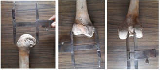
MATERIALS AND METHODS The study was conducted on 150 femur (80 right and 70 left) obtained from Department of Anatomy of a private medical college. Inclusion criteria: Intact bones with completely ossified epiphyses. Exclusion criteria: Bones with deformity, fractures, unfused epiphyses, damaged bones. Measurements were taken using a sliding calipers. Each measurement was taken twice by the same observer to reduce intraobserver bias. The results were tabulated and analysed using descriptive statistics. Antero-posterior length: anteroposterior length of the medial (APMC) and lateral condyle (APLC). Bicondylar width: maximum distance between the two femoral condyles at the level of epicondyles as shown in Figure 1. Width of intercondylar notch: maximum width of the femoral intercondylar notch as shown in Figure 2. The results were analysed using unpaired t test. The p-value of less than 0.05 was considered as statistically significant. Figure 1 Figure 2 Figure 3 Figure 1: Measurement of Anteroposterior Diameter of Condyles of femur; Figure 2: Measurement of Bicondylar width at the distal end of femur; Figure 3: Measurement of Intercondylar notch width.
RESULTS Morphometric measurements were done on 150 unpaired femurs in dry bones; out of which 80 were right sided and 70 were left sided. The results obtained of our study is shown in Table 1.
Table 1: Dimensions of distal femur
There was no statistically significant differences in the parameters between the right and left side (p value > 0.05).
DISCUSSION The mean anteroposterior lengths of medial condyle and lateral condyle observed in our study are similar to the findings of Stephen et al., Rajan et al., Biswas5,6,8 et al. as shown in Table 2. The results of the present study are higher when compared to the observations of Selvapriya et al.7 Table 2
Previous studies have shown that differences of size (anteroposterior and mediolateral width of the femur and tibia) and shape (tibial and femoral aspect ratios) exist among white, East Asian, and black populations.10 A study on anthropometric measurements of distal femur on Malay population using CT scans showed that the observed values of AP and ML for males were significantly higher than that of females. An overall comparison between the different races demonstrated that the Chinese have the largest AP and ML measurements followed by the Malay and Indian populations.11 Bicondylar width observed in the present study are close to the results of Rajan et al., Biswas et al.[6][8] Previous studies have shown that bicondylar width correlates with femoral length and has application in estimation of stature and has an important role in forensic anthropology.12 Intercondylar notch width observed in the present study is similar to the values obtained in the studies of Rajan et al., Selvapriya et al., Biswas et al., Ravichandran et al.6,7,8,9 Wada et al. suggested that a narrowing of the notch occurs after the onset of OA, leading to ACL damage, knee instability, and disease progression.13 Shepstone et al. studied the shape of intercondylar notch in arthritic and non-arthritic bone samples and suggested that difference in the shape of the intercondylar notch is a risk factor for knee OA.14 Ravichandran et al. studied the intercondylar notch dimensions and shape in dry bones and cadaveric knees and inferred that stenotic notches may be a cause for dysfunctional ACL and in extreme cases may lead to tear of the same.9 The limitations of the present study is the small sample size. However, this study provides the range of distal femur dimensions which would be useful as a reference to design knee implants suited to South Indian population.
CONCLUSIONS In the present study, mean Bicondylar width noted in the present study is 72.63 ±4.13 mm on the right side and 71.25±3.14mm on the left side. The intercondylar width observed is 21.27±4.18mm on the right side and 20.35±2.14mm on the left side. There was no statistically significant differences observed in the values of anteroposterior length of medial and lateral condyles, bicondylar width, intercondylar width between right and left sides.
REFERENCES
Policy for Articles with Open Access
|
|
|||||||||||||||||||||||||||||||||||||||||||||||||||||||||||||||||||||||||||||||||||||||||||||||||||||||||||||||
 This work is licensed under a Creative Commons Attribution-NonCommercial 4.0 International License.