 Home
Home
|
Table of Content - Volume 19 Issue 2 - August 2021
Variant muscle on dorsal aspect of hand with review of literature
Sushma Rao Poleneni1*, Urmila Sinha2, Mrudula Chandrupatla3
1Senior Resident, 3Additional Professor & HOD, Department of Anatomy, All India Institute of Medical Sciences, Bibinagar, Telangana, INDIA. 2Assistant Professor, Department of Anatomy, All India Institute of Medical Sciences, Deoghar, INDIA. Email: sushma.poleneni@gmail.com
Abstract Background: Variations in the extensor compartment of the forearm have been frequently reported as they are valuable for clinician in pre-diagnosing and planning the treatment on dorsum of hand. In one of the left upper limbs, a variation was identified where classical extensor indices muscle was completely absent and, in its place, an extra muscle, taking thin tendinous origin from upper one third of posterior surface of ulna, carpal bones, extensor retinaculum was present and it transformed into a small muscle belly which is attached distally to the dorsal digital expansion of index finger. Knowledge about rare variations helps surgeons to avoid any hand injuries, as the muscles on dorsum of hand are superficial, also it helps them during tendon transfers and muscle grafts. keywords: Extensor; Extensor Digitorum Brevis Manus; Hand; Variant; Muscle.
INTRODUCTION The extensor groups of forearm muscles help in extension of wrist and are common muscles to show variation. The synergistic contraction of extensors and flexors is mandatory for efficient grip on different objects in daily life.1 Even though variation in extensor compartment is common, they are not identified as there are no symptoms. Anomalies in origin and insertion of Extensor indices proprius and Extensor digitorum brevis manus occasionally cause symptoms which become difficult to diagnose if it is related to any other pathology.2 Clavero et al.3 used magnetic resonance imaging and showed the insertion of muscle and retinacular structure in extensor compartment. They pointed the importance of understanding the variation and normal anatomy of extensor compartment of forearm as mandatory for radiologist in diagnosing and assessment of clinical condition with MRI. The incidence of muscle variation was found to be 2% in earlier studies by Yammine K4 The extensor indices muscle takes origin from posterior surface of ulna distal to extensor pollicis longus and from the adjacent interosseous membrane. It passes in the fourth compartment below extensor retinaculum sharing common synovial sheath with extensor digitorum and lies medial to extensor polices longus. Close to head of second metacarpal bone it joins with the index tendon of extensor digitorum and enters in formation of dorsal digital expansion of index finger. This muscle was first described and named as extensor brevis digiti vel medii by Bernhard Albinus5 and the term extensor digitorum brevis manus (EDBM) was first used by Macalister.6
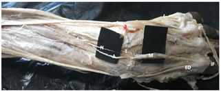
CASE REPORT A variation was observed in the extensor compartment of the left upper limb of an old male cadaver (Fig 1). The normal extensor indices muscle was absent, in its place, a small muscle belly taking origin from upper surface of carpal bones and extensor retinaculum was present. It is taking an extra tendinous origin from the posterior surface of upper one third of ulna and interosseous membrane, which is attaching to the muscle belly. The muscle along with its tendinous origin are passing in the fourth compartment along with the extensor digitorum muscle and sharing a common compartment. The muscle belly is continuing forward on dorsum of hand and getting inserted as tendon on the base of proximal phalanx of left index finger medial to extensor digitorum tendon and into dorsal digital expansion. The total length of muscle including the origin from ulna is 17.5cm. The width of muscle belly in the middle is 0.8cm, towards insertion is 0.5cm, and towards origin at extensor retinaculum is 0.6cm. The measurements of muscle are documented in a table. (Table 1) There is a separate arterial and nerve supply to the muscle arising as a branch from posterior interosseous nerve and branch of anterior interosseous artery. The posterior interosseous nerve is giving muscular branches to adjacent muscles and continuing as thin branch to supply the muscle belly. The anterior interosseous artery after piercing the interosseous membrane is giving a branch to the muscle belly (Fig 1). The dorsum of hand on right side has no variation and has a normal extensor indices muscle.
Table 1: Measurement in Centimeters of the Variant Muscle.
Figure 1: Left Upper Limb showing Extensor Digitorum Brevis Manus with Separate Nerve and Vessels [ N - Nerve; A - Artery; M - Muscle belly; T- Tendon of muscle belly, IF – Index Finger].
DISCUSSION Extensor compartment is well known for its variations, especially extensor indices proprius muscle with its origin and insertion into index and middle finger (extensor digitorum brevis manus and extensor medii proprius). Anomalous extensor tendons to fingers could be the result of regression, retention or reappearance of the changes that happen in muscles during its ontogenic development.7,8 It has been observed that most of variations in the posterior compartment were in the deep rather than in the superficial compartment as deep compartment is considered embryologically unstable. Ogura, et al.9 classified the different variants according to their insertion in relation to Extensor indices proprius muscle (EIP). They were divided into three groups: group 1 had a missing Extensor indices proprius muscle (EIP) with the Extensor digitorum brevis manus (EDBM) attached to its index finger on the dorsal aponeurosis; group 2 had an EDBM attached to the index finger along with the EIP; and group 3 had it attached to the long finger, aiding solely in extension of finger. The results of the present study indicate that the variations are closely resembling group 1 and group 3 as per the classification of EDBM by Ogura et al. In this case report, the muscle was taking extra tendinous origin from proximal one third of posterior surface of ulna and interosseous membrane, also from the posterior surface of carpal bones and extensor retinaculum. It is sharing a common compartment with extensor digitorum, so it might lead to symptoms of compression of nerve and artery. The measurements of the muscle are tabulated, which further help surgeons during tendon grafts, tendon transfers in the extensor compartment. The reason for the variation may be embryological or hereditary factor. Reports suggest that it represents the failed proximal migration of the undifferentiated extensor muscle mass of hand similar to an atavistic structure10 Study by Gama C11 reported the existence of familial dominance of EDBM muscle which is still a matter of debate. Peculiarity of this variation is that most of the cases were identified during dissection or surgeries but few are pre diagnosed with the presence of symptoms like chronic pain on dorsal aspect of hand during extension12 Pain is usually aggravated by hand dominance and manual labor. For clinician to not to miss out diagnosis, knowledge of variation is crucial factor. The fourth compartment of extensor region has four extensor digitorum tendons crowded over extensor indices tendon along with posterior interosseous nerve and anterior interosseous artery. According to McMinn an extra tendon can cause increase pressure in this rigid Osseo fibrous tunnel and leads to compression of the muscle fibers and the posterior interosseous nerve, which leads to fourth compartment syndrome.13 Hayashi, et al.12 coined the term “Fourth Compartment syndrome” which reasoned the chronic dorsal wrist pain to etiology in extensor retinaculum. Further, they listed the five possible causes as EDBM, abnormal extensor indices, ganglion presence, tenosynovitis and abnormal carpal bones. Patel, et al.14 proposed that the first treatment should be retinacular release, especially when EDBM serves to compensate for the EIP. Waterman et al.,15 said that surgical excision was more helpful than debridement or retinacular release. Waterman conducted an operation where the muscle was misdiagnosed as scaphoid pathology and later diagnosed as EDBM. More researchers should evaluate the variations and improve imaging technique to diagnose the condition and develop the treatment mode. We conclude with the statement that EDBM has evaded diagnosis in many patients so, there should be a proper imaging techniques and knowledge about the origin, insertion, nerve and arterial supply of muscle. In this case, the anomalous muscle resembles EDBM, has separate arterial and nerve supply with extra tendinous origin from proximal one third of ulna. In most of the cases extra muscle on dorsum of hand are not diagnosed as they are asymptomatic but in athletes like weight lifters, tennis players, cricketers and also in manual laborers due to repeated extension it might lead to symptoms. So, such rare variations should be documented as they help surgeons to avoid diagnostic error and help in surgical intervention. Further studies on familial dominance should be considered for better diagnosing and treating the condition.
REFERENCES
Policy for Articles with Open Access
|
|
 This work is licensed under a Creative Commons Attribution-NonCommercial 4.0 International License.