 Home
Home
|
Table of Content - Volume 20 Issue 1 - October 2021
Variations in median nerve formation
Shobha Verma1*, Nilofer Mulla2, Manisha Nakhate3
1Assistant Professor, 3Professor & HOD, Department of Anatomy, D Y Patil Medical College, Navi Mumbai, Maharashtra, INDIA. 2Assistant Professor, Department of Anatomy, HBT Medical College & Dr. R N Cooper Municipal General Hospital, Mumbai, Maharashtra, INDIA. Email: drshobhaverma20@gmail.com
Abstract Background: Normally median nerve is formed by union of a lateral and medial roots lateral to third part of Axillary artery. A number of variations in the formation of median nerve were reported in the literature. Some authors reported the formation of median nerve by three roots, two lateral roots and one medial root. Formation of median nerve by a single root from lateral cord has also been reported. In present study we observed the cases formation of median nerve by three roots of brachial plexus. Material And Method: We studied anatomical variation in 24 embalbed human cadaver in Department of Anatomy. Out of fourty eight upper limbs dissected, three upper limbs showed variation in the formation of median nerve. During routine cadaveric dissection we noted the formation of median nerve by three roots. Result: In a male cadaver on the right upper limb, median nerve was formed by three roots, one lateral and a medial root joining to form median nerve lateral to axillary artery and a second lateral root piercing through coraco-brachialis muscle joining median nerve at a distal level. An anomalous profunda brachii artery arising from third part of axillary artery was also noted passing between the two roots of median nerve. In another cadaver on the left upper limb, median nerve was formed by three roots, two lateral and one medial with one of the lateral root joining distally. In third cadaver on the right side, median nerve was formed by two medial roots and one lateral root. All the varation were unilateral Conclusion: These variations are clinically important for surgeons, anaesthetist and orthopedician.
INTRODUCTION Median nerve is a branch of brachial plexus which is formed by the two roots. Lateral cord gives rise to lateral root of median nerve, while medial cord gives out medial root of median nerve. Normally the median nerve is found lateral to the third part of axillary artery by the union of medial and lateral roots. Formation is “Y” shaped and medial root crosses in front of the third part of the axillary artery.8 A number of variation of the formation of median nerve formation and its relationship have been reported by earlier workers. Knowledge of median nerve formation and its variable relationship to the axillary artery is important during surgeries in axillary region , anaesthetic procedure of brachial plexus ,shoulder arthroscopy and traumatic injuries involving axillary region. Injury of such variant may lead to galaxy of complications.
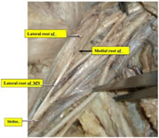
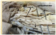
MATERIAL AND METHOD During routine cadaveric dissection in the department of Anatomy anamolous median nerve with regard to their formation is found in three different cadaver out of 24 embalbed cadaver or 48 upper limb . All these variations were unilateral . OBSERVATION Following variation related to the formation of median nerve were observed. CASE 1: In middle aged female cadaver on right side median nerve was formed by 3 roots.Two roots were coming from the lateral cord ,and one root from the medial cord.The extra root coming from the lateral cord pierces the corachobrachialis muscle and join the median nerve distal to the insertion of pectoralis major.Musculocutaneous nerve has its normal course through the corachobrachialis.accroding to Venierators et al. (1998)3 there were three types of communication between musculocutaneus nerve and median nerve . Type 1: Communicaion of musculo-cutaneous nerve and median nerve proximal to the entrance of musculo-cutanoeus nerve into coracobrachialis. Type 2: Communicating trunk passes through coracobrachialis to join median nerve. Type 3: musculocutaneous nerve and communicating trunk did not pass through coracobrachialis muscle. In the present case the communicating trunk passes through coraco-brachialis muscle to join the median nerve hence it is Type 2 Figure 1 CASE 2: In a aged male cadever on left side median Nerve is formed by 3 roots; 2 lateral and one medial root. One of the lateral root arised from the musculocutaneous nerve and other directlt from lateral part. The communicating branch from the musculocutaneous nerve is type 1 because it is arising from musculocutaneous nerve before its entry into coraco brachialis muscle. Figure 2
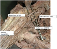
CASE 3: In another aged male cadever on right side median nerve was formed by 3 roots; 1 lateral and 2 medial roots. The smaller medial root joined more proxmally and the larger one distally. Median nerve lied medial to the axallry artery because the lateral root crossing in front of 3rd part of Axiallry artery to join the medial root. There are no previous reported cases of presence of 2 medial roots. Figure 3
RESULT The present study done on 24 embalbed cadavers ( 48 upper limb) out of which 3 upper limb shows variation in median nerve root formation .In two cases median nerve is formed by one medial root and two lateral roots. In third case median root is formed by two medial and one lalteral root.
DISCUSSION Median Nerve formation by 3 roots: Variation in the median nerve was demonstrated by some past workers. However most of the variation noted by them were related with formation of median nerve by 3 roots. Sargon et al. ( 1995)1 reported a case of 55 year old male in which median nerve had an additional contribution from lateral cord.Both branches from the lateral cord crosses the axiallry Artery anteriorly and joined the medial root medially to the axiallary artery. Abhaya et al. ( 2003)7 reported a case of 80 year male cadever in which lateral cord pierced the coracobrachialis muscle from its medial side. 2 branches arising from the anterior aspect of the coraco bracialis muscle to joined the medial root of the median nerve. Pandey and shukla reported a case in( 2006),10 in which median nerve formed by contribution of all 3 cords of brachial plexus. Chitra et al. (2007)9 studied 50 upper limbs of which 13 upper limbs showed formation of median nerve by 3 roots. In all cases the 3rd branch arised from musculocutaneous nerve. Anyanwu et al. ( 2009)12 studied 50 upper limbs of which 3 uppe r limbs showed formation of median nerve by 3 roots. 3rd root arised form musculocutaneous nerve. Satyanarayan et al. ( 2009)13 reported a case in which median nerve formatiion by 3 roots , 2 from lateral and one from medial cord. In this case median nerve was formed anterior to the axillary artery. Buddhiraja et al. (2009),15 studied 196 upper limbs.He noted 3 roots were taking part in formation of the median nerve. Out of 196 upper limb that he studied, in 28 upper limb 3rd root arised from lateral cord and in 16 upper limbs 3 rd root arised from musculocutaneous nerve. Talhar et al.( 2012),16 reported a case in which there was formation of median nerve by 3 roots, 2 roots from lateral cord and one from medial cord. Apurva et al.( 2013),17 studied 100 upper limb of which 3 upper limb showed median nerve formation by 3 roots and 3rd root coming from lateral cord. In 6 cases 3rd root was coming from musculocutaneous nerve before piercing the coraco brachialis muscle which is further supplied by median nerve. Sadananda rao et al. 201318 reported a case of median formation by 3 roots, in which 3rd root was arising from lateral cord. Eglseder and Goldman (1997)2 have found that median nerve was formed from three roots two lateral and one medial root in 14% of specimens Chauhan and Roy (2002),5 Satyanarayana et al. (2010)14 and Saeed and Rufai (2003)8 also observed the same variation Median nerve formation by 4 roots Badawoud (2003)6 showed 4 types of anomalies in the median nerve formation found on 48 upper extremities of 24 cadaver anomalies (usually on the left side) which comprised of communicating branches between lateral and medial roots of the median nerve, a communicating branch between lateral root of the median nerve and the medial cord of the brachial plexus, communicating branches between lateral root of the median nerve and musculocutaneous nerve and unusually long roots of the median nerve Uzun and seeling in( 2001)4 reported a case of 66 year old male cadever . In this case the median nerve was formed by 4 roots. 3 from lateral cord and one from medial cord. Satayanarayan et al.( 2009)13 reported that median nerve was formed by 4 roots. 3 from lateral cord at diffreent level: (i) Upper at the level of coraco brachialis. (ii) Middle one below coraco brachialis. (iii) 3rd at the upper border of insertion of lattissmus dorsi muscle. Dwarish etal in( 2009)11 reported a case of median nerve formation from 4 roots, 3 from lateral cord and one from medial cord. Buddhiraja et al. (2010)15, studied 196 upper limb n which 7 upper limb shows median nerve formation by 4 roots. 3rd and 4th root from lateral cord and musculocutaneous nerve. The knowlege of these variations is importantssss to the surgeon for carrying out surgical procedures related to axilla and arm. Because while performing surgeries in these region injury to such variant forms can lead to various manifestations including sensory, motor or vasomotor or trophic changes.
REFERENCES
Policy for Articles with Open Access
|
|
 This work is licensed under a Creative Commons Attribution-NonCommercial 4.0 International License.