 Home
Home
|
Table of Content - Volume 20 Issue 3 - December 2021
Cadaveric study on the variations in the origin, course, pattern and termination of the azygous venous system
Ramya P P1*, Lakshmikantha B M2, Sukritha K M3
1-3Assistant Professor, 2Professor, Department of Anatomy, DM Wayanad Institute of Medical Sciences, Meppadi, Wayanad, Kerala-673577, INDIA. Email: sukrithakm86@gmail.com Abstract Background: The Azygous system of veins are unpaired veins draining the blood from the chest, abdominal walls and the back. The variant Azygous vein can cause difficulty in diagnosing the structures in magnetic resonance imaging scans and also during surgeries involving these areas. There is a need to study the variations in the azygous system. Aim and objectives: To study the origin, course, pattern and termination of the Azygous, Hemiazygous and Accessory Hemiazygous veins. Materials and method:This study was conducted in the Department of Anatomy, DM WIMS Medical college, Wayanad, Kerala from 2018 to 2021 using 25 formalin fixed dissected thoracic specimens. Observation and result: Among the 25specimen studied the azygous originated from the lateral root (right ascending lumbar and right subcostal veins) in all cases. The Azygous mostly followed a midline course (40%) and terminated into the superior vena cava at the level of 3rd thoracic vertebra (56%). The Hemiazygous and Accessory Hemiazygous veins were present in 84% and 68% respectively where 66.6% of Hemiazygous terminated at the 8th thoracic vertebral level and 82.3% of Accessory Hemiazygous at 7th thoracic vertebral levels. In the study, the azygous system showed type II variant (92%) and subgroup 2 pattern was seen in 20% cases. Conclusion:Surgeons and radiologist should have a clear knowledge about the variations in the Azygous vein. This will help to avoid miss diagnosis and accidental injury to this vein leading to complications. Key words: Azygous vein, Hemiazygous vein, Accessory Hemiazygous vein,
INTRODUCTION The Azygous system is the unpaired venous system1 consisting of the Azygous vein, the inferior Hemiazygous (Hemiazygous) and the superior Hemiazygous (Accessory Hemiazygous vein). They drain blood from the posterior part of the chest, posterior abdominal wall and related structures. The Azygous vein is formed by the union of the right ascending lumbar and the right subcostal vein. It passes upwards along the right of the thoracic vertebral bodies, arches at the level of 5th thoracic vertebra and drain into the superior vena cava. The tributaries of the Azygous vein include the right superior intercostal, lower right posterior intercostal, superior and inferior hemiazygous and mediastinal veins. The Hemiazygous is formed by the union of the left ascending lumbar and the left subcostal vein. It drains into the Azygous at the level of 8th thoracic vertebra. Lower left subcostal and the mediastinal veins are the tributaries of the hemiazygous vein. The 4th to 8th left posterior intercostal veins join to form the inferior (accessory) hemiazygous vein. It drains into the Azygous vein at the level of 7th thoracic vertebra.2 There are numerous variations in the Azygous system. The Azygous may originate from a combination of lateral, intermediate or medial roots. The lateral root is formed by the union of the right ascending lumbar and the right subcostal. The intermediate root arise from the dorsal aspect of the inferior vena cava and the medial root arise as plexiform complex from lumbar segmental branches.3 The Hemiazygous and the Accessory Hemiazygous may be rudimentary or absent. In such cases the left posterior intercostal veins will directly drain into the Azygous vein. Various anastomoses exist between the tributaries and the main azygous vein. There can be connections between the Hemiazygous and Accessory Hemiazygous vein also. The variations in the Azygous system is closely related to its developmental aspects. The Azygos vein develops from the upper right supra cardinal vein and the Azygos arch from the upper segment of the right posterior cardinal vein.4 The 4th to 11th right intercostal veins end in the right supra cardinal vein which is joined with a portion of right posterior cardinal vein to form the Azygos vein. The 4th to 11th left intercostal veins end in the left them at 6th and 7th thoracic vertebrae in adults. The left supra cardinal vein which develops into the Hemiazygos vein that ends into the azygos vein.5 A transverse communication is formed between upper part of this anastomoses forms the Accessory Hemiazygous vein. The Azygous vein may sometimes be present in the midline or towards the left of the midline anterior to the vertebral bodies.7 The variation in the course may arise due to degenerative or pathological changes during ageing process, one of the causes being osteophytosis along the vertebral column (it is caused by the mechanical stresses and strains on the vertebral column).8 The anatomical variations in the Azygous vein should be known to the surgeons and the radiologist. These structures can be misinterpreted as an aneurysm, enlarged lymph node or tumour in CT and MRI scans.9 Surgeons should have knowledge of these variations during operations in mediastinum to avoid severe bleeding. Thromboembolic diseases in young can arise from these congenital venous abnormalities.10 Some of the surgeries for the trauma care may require ligating the inferior vena cava that can follow this system, hence any abnormal course should be known to the surgeons to avoid complications.11 The aim of the present study is to determine the variations in the origin, course, branching patterns and termination of the Azygous, Hemiazygous and the accessory Hemiazygous veins.
MATERIALS AND METHOD This study was conducted in the Department of Anatomy, DM WIMS Medical college, Wayanad, Kerala from 2018 to 2021 using 25 formalin fixed dissected thorax specimens. The variations in the formation, course, pattern, level of termination of the Azygous vein along with the presence and level of termination of Hemiazyous and Accessory Hemiazygous vein were noted and analyzed using Excel sheet. The pattern of the Azygous system was noted. The specimen were numbered and photographs taken.
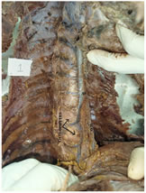
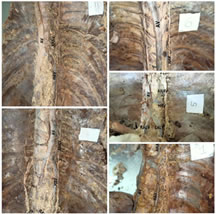
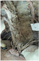
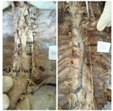
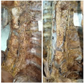
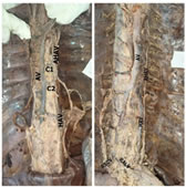
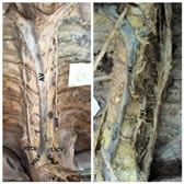
OBSERVATION AND RESULT In the 25specimen studied, the Azygous vein formation was noted from the lateral root, by the union of the right ascending lumbar and the right subcostal veins in all the cases (100%). The course of the Azygous vein studied in the 25 specimen showed that the Azygous followed a midline course in 40% cases, among which few were attributed to the presence of the osteophytes(figure 1). The Azygous had a right sided course in 24% and left sided in 36% cases. It was noted that the Azygous vein terminated at the level of 3rd thoracic vertebra in 56% cases followed by 40% at 2nd thoracic and 4% at 4th thoracic vertebral levels. The Hemiazygous and Accessory Hemiazygous veins were present in 84% and 68% respectively. The Hemiazygous vein terminated at the level of 8th Thoracic vertebra in 66.6%, 7th thoracic vertebra in 23.8% and 4.7% each at the level of 9th and 10th thoracic vertebrae. In 82.3% cases the Accessory hemiazygous vein terminated at the level of 7th thoracic vertebra. 5.8% cases each were noted to terminate at the level of 5th, 8th and 9th thoracic vertebral levels. The patterning of the Azygous system was done as per that of Anson et al.12 who classified the Azygous into 3 types and 11 subgroups. In the study 92% of cases showed type II pattern, transition type (figure2 shows different variants of type II) and 8% showed type III variant that is with a single azygous vein in the midline (Figure 3). Type I (primitive) was not observed in the present study. The type II pattern was divided into subgroups 2 to 10 based on the number and arrangements of the communication channels. In the present study, the subgroup 6(Figure 4) was seen in 24% cases, subgroup 2(Figure 5) in 20% cases, subgroup 4 in 16%. 12% cases showed subgroup 3 and subgroup 4 each and 8 % each had subgroup 5(Figure 6) and subgroup 10(Figure 7). The subgroups 7, 8 and 9 were not observed in the present study.
DISCUSSION The formation of the Azygous vein was noted to be from the lateral root (right ascending lumbar and right subcostal veins) in all the 25 cases studied (100%). This finding was corresponding to the findings from other studies done by Kanchana et al.,13 George AS14 and Bergman.3 Kanchana et al.13 had stated that among the 100 specimen studied, the Azygous vein formed from the lateral root in 100% cases. George AS14 and Bergman3 had noted the same in 94% and 85% cases respectively. According to Elizbieta Krakow15 the Azygous vein had a right sided course in 90.6% and midline course in 9.4%, Tatar et al.16 noted that the Azygous vein was in the right in 37.9%, 22.3% in left and 39.85 in the midline and Nathan17 noted that the Azygous was in the right in 20%, left in 53% and midline in 27%. In the present study, the Azygous vein followed a midline course in 40%, left side in 36% and right side in 24% which were comparable with that of the studies by Tatar et al.16 According to Kutoglu6 the Azygous vein terminated into the superior vena cava at the level of 3rd thoracic vertebra(85%).
Figure 1 Figure 2 Figure 3 Figure 4 Figure 5 Figure 6 Figure 7 Figure 1: Midline course of the Azygous vein due to the presence of osteophytes; Figure 2: Different subgroups of Type II pattern; Figure 3: Azygous venous system with Type III pattern; Figure 4: Azygous system with type II subgroup 6 pattern; Figure 5: Azygous system with type II subgroup 2 pattern; Figure 6: Azygous system with type II subgroup 5 pattern; Figure 7: Azygous system with type II subgroup 10 pattern.
The present study had also showed that in majority of cases (56%) the Azygous vein terminated at the level of 3rd thoracic vertebra. The result had differed from that of Tatar et al.16 which showed termination between 5th and 6th thoracic vertebral levels and Kanchana13 noted the termination at the level of 4th thoracic vertebra in 85% cases. The Hemiazygous vein terminated at the level of 8th thoracic vertebra in 66.6% cases in the present study which were in comparison with that of the studies done by Elzbieta15 which showed 35.7% cases terminated at the level of 8th thoracic vertebra, Kutoglu6 showed 27.1% and Kanchana13 showed 70% terminated at the level of 8th thoracic vertebra. The termination of the Accessory hemiazygous vein was noted to be at the 7th thoracic vertebral level in 82.3% cases which was similar to that seen by Elzbieta,15 which showed 41.6% cases. The pattern of the Azygous venous system was widely studied by many authors. Anson12 had classified the Azygous system into 3 types and 11 subgroups. According to him, Type I was seen in 1%, Type II in 98% and type III in 1% cases. According to Kutoglu6 2.1%, 91.7% and 2.1% cases were seen respectively in type I, type II and type III patterns. Dahran and Soames18 noted type I in 3%, type II in 87% and type III in 10% cases. George AS14 had noted type III pattern in 5% cases. The same classification had been used in the present study also. It was found that 92% showed type II pattern and 8% showed type III pattern which were comparable with other studies like Anson.12 Kutoglu6 and Dahran and Soames.18 Type 1 pattern was not noted in the present study.
CONCLUSION The variations in the Azygous system have been studied and analysed. The knowledge about the variation in the Azygous system is highly important for the surgeons and the radiologist while assessing a patient for surgical interventions. The variations in the Azygous venous system may be confused with aneurysm and other abnormalities like tumour and lymphadenopathy. It is important to have knowledge of these variations while performing the mediastinal operations or surgery of large vessels. Any deviation from the normal state can cause a treat to the surgeons. A thorough knowledge will aid them in easy diagnosis as well as minimize the complications during the surgeries. The variations noted in the cadaveric dissection can be used as an aid to visualize the Azygous system in the computed tomography and magnetic resonance imaging. The knowledge of the anatomical variations may aid in detecting the abnormalities and can be used as future aids during the procedures involving the same.
REFERENCES
Policy for Articles with Open Access
|
|
 This work is licensed under a Creative Commons Attribution-NonCommercial 4.0 International License.