|
|
|||||||
|
|
|||||||
 |
|||||||
|
|
[Abstract] [PDF] [HTML] [Linked References]
A Rare Combination
Hina Kauser1, Waseem Anwar2, Maniah Qadir3 1Assistant Professor, 2,3Senior Resident, Department Of Ophthalmology, Hamdard Institute of Medical Sciences and Research, Jamia Hamdard University, Hamdard Nagar, New Delhi -110062 INDIA. Corresponding Addresses: 1hina3kauser@yahoo.co.in, 2muneeb2rehman@hotmail.com, 3qadirmaniah@gmail.com Case Report
Abstract: Primary angle closure (PAC) usually occurs in hypermetropic eyes, which have a short axial length and a shallow anterior chamber. In myopic eyes, the axial length is usually longer than average and the anterior chamber is usually deep; PAC is rarely observed in these eyes. We are reporting a case of 40 years of female presenting to our hospital with pain and decreased vision in one eye. The patient was diagnosed to have an acute attack of angle closure glaucoma. Investigations showed the patient to be a high myope of -14.5 D. Only few cases with acute angle closure glaucoma associated with high myopia have been reported and to my knowledge this is the youngest and most myopic patient reported till date in India having angle closure due to pupillary block. The case report of this patient illustrates that myopia does not exclude the presence of angle closure and this should be sought by gonioscopy by ophthalmologist and general physicians should also suspect that angle closure glaucoma could be one of the cause of red painful eye even though the patient is a known high myope. Keywords: high myopia, primary angle closure glaucoma, gonioscopy.
Introduction Narrowing of anterior chamber angle and angle closure glaucoma usually occurs in hypermetropic eyes. In primary angle closure glaucoma (PACG) the most common cause of angle closure is pupillary block and elevation of intraocular pressure occurs as a result of obstruction of aqueous outflow by partial or complete closure of the angle by the peripheral iris. The relative pupillary block is typically found in eyes having hypermetropic refractive error, shallow anterior chamber depth, short axial length and larger lens thickness. Hypermetropic eyes relatively having short axial length and small corneal diameter are at increased risk of PACG. [1], [2] Myopia is observed rarely in patients with these conditions as the myopic eyes typically have deep anterior chamber and wide opened anterior chamber angles, these anatomical factors associated with myopia offer considerable protection from angle closure glaucoma, but inspite of all these ,the presence of myopia does not eliminate the possibility of primary angle closure glaucoma. We report the patient presented with acute angle closure glaucoma associated with -14.5D myopia. Laser peripheral iridectomy was done to relieve the relative pupillary block.
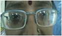
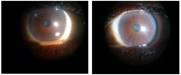
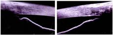
Figure 1: Patient wearing high Figure 2: laser PI done BE Figure 3: Ant seg OCT showing narrow angle BE power glasses
Case History A 40 years female presented in our Hospital with blurring and pain in her right eye for 1 day. No history of any medicine intake and trauma to eye was there. She had the history of wearing thick glasses since her childhood. [Figure 1] Her family history was unremarkable. Visual Aquity in right eye was 20/200 and in left eye was 20/40 with glasses having refractive status of -14Dsph -1 D cyl axis180 in right eye and-13D sph -1.25cyl axis 170 in left eye. Slit lamp examination revealed circumciliary congestion, hazy cornea with epithelial oedema, shallow anterior chamber with mid-dilated pupil. slit lamp examination of left eye was normal except for shallow anterior chamber( grade 2 - van Herrick’s).Intraocular pressure was 58mmHg in right eye and 16 mmHg in left eye with applanation tonometry.A diagnosis of acute angle closure glaucoma was made on the basis of clinical findings . Patient was managed by giving intravenous mannitol 300ml, acetazolamide 500mg orally, pilocarpine eye drop every 15 min and timolol eye drop initially. Patient was examined after 2 hours; there was decrease in pain and redness, relative decrease in corneal haziness, decrease in dilatation of pupil .Intraocular pressure reduced to 22 mmHg. Gonioscopy was done and findings were grade 0-1 in inferior quadrant, grade 0 in rest 3 quadrants in right eye and grade 1 in superior and temporal, grade 2 in rest 2 quadrants (Shaffer’s grading).No iris processes and peripheral anterior synechia seen on gonioscopy. YAG laser peripheral iridectomy was performed in both eyes subsequently [Figure 2].Post laser peripheral iridectomy pupil was dilated and proper examination was done to rule out lens subluxation, spherophakia, iris cysts .Fundus examination revealed tessellated fundus with optic disc showing 0.3 cupping .A-scan revealed axial length of 26.3 and 25.9mm in right and left eye respectively, lens thickness of 4.78 and 4.58mm in right and left eye respectively and anterior chamber depth was 2.07 mm in right eye and 2.37 in left eye. Anterior segment optical coherence tomography showed shallow anterior chamber with no pathology in angle and iris. [Figure 3]. Corneal diameters was 11.2 mm (horizontal) and 11mm (vertical) in both the eyes and measured with calipers. Discussion This case demonstrates the uncommon presentation of angle closure glaucoma in highly myopic middle aged female. The most common mechanism of angle closure is pupillary block .The typical eye with primary relative pupillary block is hypermetropic eye and angle closure with relative pupillary block typically occurs in older individuals as continuous growth of lens throughout adulthood results in increased lens thickness with decrease in anterior chamber depth and volume. Narrow angles can also occur secondary to iris cyst, plateau iris ,lenticular abnormalities, certain medications which were all absent in our case .In literature very few cases have been reported of angle closure glaucoma due to primary pupillary block associated with high myopia . Lowe reported that out of 127 eyes of PACG patients, only 2 had myopia in the range of -6.25 to -8 D. [2] Marchini G et al carried out biometric study in 54 patients of primary angle closure glaucoma, all the 54 patients were showing smaller than average axial length, in the range of 22.31+_0.83mm confirming it is a disease of small hypermetropic eyes. [3] Hagan and Lederer reported the most myopic patient of -8.62 D was associated with PACG but subsequent observations after 7 years has revealed that angle closure glaucoma was due to ectopia lentis.[4],[5] Barkana reported 20 myopic patients in a series of 17, 938 examined over 20 years having narrow angle glaucoma with only 9 myopic patients having angle closure because of primary pupillary block and in rest 11 a spectrum of ophthalmic conditions lead to myopia and angle closure and most myopic patient in primary pupillary block group was having -13.25 D and youngest patient of primary pupillary group was 52 years of age. [6] A study in 692 patients of angle closure was done in china in 2009 only 5 patients have myopia with angle closure due to primary pupillary block.[7] Nancy reported a case of narrow angles with high myopia of -8.5 D in which spherophakia is the cause of narrowing of angles and lenticular myopia.[8] 322 cases of primary angle closure glaucoma were reviewed by Chakravarti T, only 6 cases occurred in myopic eyes.[9] Shaukat Ali reported a case of angle closure in a myope of less than -6D.[10] This shows that high myopia is rarely associated with angle closure glaucoma but this does not exclude the presence of narrow angles and that this should be sought by gonioscopy. We advocate ophthalmologist to perform careful gonioscopy in all patients undergoing initial examination irrespective of refractive status and general physicians should also suspect that angle closure glaucoma could be one of the cause of red painful eye even though the patient is a known high myope. References
|
||||||
|
|||||||