|
|
|||||||
|
|
|||||||
 |
|||||||
|
|
[Abstract] [PDF] [HTML] [Linked References]
Pregnancy in non Communicating Horn of Uterus – A Case Report
Shreyaa Sriram1*, U. T. Bhosale2, Ramchandra D. Shriwastav3 1PG student 1st year, 2HOD, 3Assistant Professor Department of Obstretics and Gyanaecology, Bharati Vidyapeeth Medical College and Hospital, Sangli, Maharashtra, INDIA. *Corresponding Address: Case Report
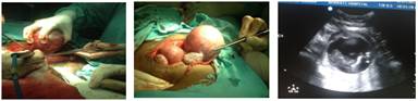
Abstract: The incidence of uterine malformations is estimated to be 3% - 5% in the general population. Abnormal fusion of the mesonephric duct (mullerian duct) during embryonic life results in a variety of congenital uterine malformations like bicornuate uterus, unicornuate uterus. A unicornuate uterus is a uterus that has a single horn and a banana shape. Approximately 65% of women with a unicornuate uterus also have a second smaller or rudimentary uterine horn. The rudimentary horn can be solid or it can have a small cavity with a functioning ndometrium. Sometimes the smaller horn connects to the uterus and vagina, but more often it is isolated or non-communicating. Pregnancy can also occur in a non-communicating arm. The situation is similar to an ectopic pregnancy and must be treated as an emergency. If pregnancy occurs in the non-communicating arm, uterine rupture occurs in approximately 89% of cases by the end of the second trimester. Because of this risk, surgical removal of the non-communicating arm is recommended. Removal of a solid non-functioning arm is not necessary. In this case it is presented as a 30 year old 3nd Gravida with 1st spontaneous abortion, 2nd pregnancy she underwent LSCS for breech presentation delivered 3kgs male baby, living, came with H/O 4 months Amenorrhea and P V bleeding since 2 days admitted in Bharati Hospital, Sangli. Patient was investigated with complete ANC profile and USG Obstetrics. USG was suggestive of uterus an teverted and bulky, E/O pregnancy of 13 weeks and 4 days, E/O Acrania with Exomphalous, FL -12mm was noted, Placenta attached to the fund us of the uterus. Impression -IUD with anomalies in baby with Bicornuate Uterus having pregnancy in right horn of uterus. Under ultrasonography guidance Emcridyl instillation was tried but failed due to? non communication of right horn of uterus to cervix. Decision of exploratory laprotomy was taken to terminate the pregnancy, on opening abdomen it was found that uterus was having a right horn enlarged with pregnancy of 12 – 14 weeks. Left horn, tubes and ovaries normal. Right horn with pregnancy excised. Postoperative period was uneventful. In conclusion, it will be interesting to know, if history of previous caesarean sections for breech (as observed) might be a probable etiological factor for rudimentary horn pregnancies and it is suggested that earlier detection of the location of embryonic growth by sophisticated diagnostic tools will save any such catastrophic outcome. Aim: Bicornuate uterus, also commonly referred to as a "heart-shaped" uterus, is a type of uterine malformation where two "horns" form at the upper part of the uterus. A bicornuate uterus is formed during embryogenesis. The fusion process of the upper part of the Müllerian ducts is altered. As a result the caudal part of the uterus is normal while the cephalo part is bifurcated. In this case it is presented as a 30 year old 3nd Gravida with 1st spontaneous abortion, 2nd Prev LSCS for breech presentation delivered 3kgs male baby, living came with H/O 4 months Amenorrhea and P V bleeding since 2 days admitted in Bharati Hospital, Sangli. Patient was investigated with complete ANC profile and USG Obstetrics. USG was suggestive of uterus anteverted and bulky, E/O pregnancy of 13 weeks and 4 days, E/O Acrania with Exomphalous, FL -12mm was noted, Placenta attached to the fund us of the uterus. Impression -IUD with anomalies in baby with Bicornuate Uterus having pregnancy in right horn of uterus. Under ultrasonography guidance Emcridyl instillation was tried but failed due to? non communication of right horn of uterus to cervix. Results: Decision of exploratory laprotomy was taken to terminate the pregnancy, on opening abdomen it was found that uterus was bicornuate with right horn enlarged with pregnancy of 12 – 14 weeks. Left horn, tubes and ovaries normal. Right horn with pregnancy excised. Conclusion: This was a rare and challenging case for us. This corrective surgery does not impair fertility. Women with uterine anomalies have poorer reproductive outcomes and lower pregnancy rates compared with women who posses normal uterus. Keywords: developmental anomaly, mesonephric duct, unicornuate uterus, miscarriage, pregnancies, fetus, complications.
Introduction Congenital malformations of the uterus, also known as mullerian duct anomalies, are rare in general population approx 1%. These abnormalities result from arrested development, abnormal formation or incomplete fusion of mesonephric ducts. Unicornuate uterus results from unilateral arrested mullerian duct development. Rarely unicornuate uterus may also have a rudimentary horn, more on the right than on the left side. The incidence of unicornuate uterus is estimated to be 1:250 and its occurrence with rudimentary horn is 1:100,000. Such anomalies are reported to result in increased rate of miscarriages, recurrent pregnancy losses, preterm labor, infertility and other obstetric complications. Conception in rudimentary horn arises either from a small communication with the uterine cavity (communicating) or by transperitoneal migration of the fertilized ovum from contra lateral side (non communicating). The proportion of non communicating rudimentary horns is 70-90%. The frequency of pregnancy in rudimentary horn is reported to be 1:76000. The clinical presentations vary from being asymptomatic to vague complaints of mild lower abdominal pain with gastrointestinal upset to its severest form of acute abdomen and sometimes with hemorrhagic shock. The most common threat of rudimentary horn pregnancy is the risk of rupture because of poorly developed musculature. In view of scarcity of literature on observation of pregnancy in rudimentary horn of uterus, the case reported here is of importance.
Materials and Methods The present study on a case report of pregnancy in non communicating horn of uterus was conducted in obstetric and gynaecology department of bharati hospital, bharati vidyapeeth university medical college, sangli, Maharashtra, India during period august 2013 to September 2013.
Case Report A 30 yr old lady, married for 8 years, para 1 with history of previous caesarean section presented to emergency department with complaints of PV bleeding since 2 days at 16 weeks of gestation. Her previous caesarean section was for breech presentation. She also had a history of a spontaneos abortion.Her vitals were stable. On abdominal examination, the uterus was 16 weeks size and irritable. Per speculum examination showed that the cervix was open mild bleeding through OS was present. Per vaginal examination showed that OS 1.5 cm dilated, no fetal parts felt. Respiratory and Cardiovascular system examinations had no significant findings. Ultrasonography was suggestive of uterus anteverted and bulky, E/O pregnancy of 13 weeks and 4 days, E/O Acrania and Exomphalous, FL -12mm was noted, Placenta attached to the fund us of the uterus. Impression -IUD with anomalies in baby with Bicornuate Uterus having pregnancy in right horn of uterus. Under ultrasonography guidance Emcridyl instillation was tried but failed due to? non communication of right horn of uterus to cervix. Decision of exploratory laparotomy was taken to terminate the pregnancy. During the procedure, while opening abdomen it was found that the right horn was enlarged with pregnancy of 12 – 14 weeks. Left horn was normal with a normal ovary and fallopian tube. Right horn with pregnancy and adenexa was excised. Operative findings were Resection of accessory horn of uterus was done with repair of sliced segment of uterus. Baby had acrania with exophthalous. The patient had a smooth postoperative recovery and was discharged from the hospital and was asked to come for follow up after 6 weeks during which her HSG was done which showed single horn of uterus on the left side with tube, showing the patency of the tube.
Discussion Women with uterine anomalies have poorer reproductive outcomes and lower pregancy rates compared with women who posses normal uterus. With introduction of MRI and 2D Ultrasonography, increased rate of accurate diagnosis is now possible. Obstetrical complications such as preterm delivery and 1st trimester miscarriage are higher in women with abnormal uterus. More than 50% of women with malformed uterus will stay completely asymptomatic. Obstetrics complications are infertility, early abortions, ectopic pregnancies, late abortions or premature birth, and IUGR. Embryogenesis The uterus is developed from the fused caudal vertical parts of the paramesonephric ducts, and the site of angular junction becomes the cervix dome and forms the fundus of the uterus. The fusion between the ducts is incomplete at first, a septum persisting between the lumina. Later, the septum disappears so that a single cavity remains. The upper part of the cavity forms the lumen of the body and cervix of the uterus. The myometrium is formed from the surrounding mesenchyme.Failure of the paramesonephric duct to fuse may cause a variety of uterine defects. They are
References
|
||||||
|
|||||||