|
|
|||||||
|
|
|||||||
 |
|||||||
|
|
[Abstract] [PDF] [HTML] [Linked References]
Unusual Fungus Isolated in Leg Ulcer with Swelling
Ghosh Kumarjyoti1*, Ghosh Bharati2, Roy Atanu3, Bhattacharya T. K.4, Ghosh A. J.5 M.G.M. Medical College and L.S.K. Hospital, Kishanganj, Bihar, INDIA. *Corresponding Address: Case Report
Abstract: Introduction: Chromoblastomycosis also known as chromomycosis, cladosporiosis, fonseca disease, pedroso disease or verrucose dermatitis (1) is a long term fungal infection of skin and subcutaneous tissue (2). The infection occurs most commonly in tropical and subtropical climates obtained in rural areas. It can be caused by many different types of fungi which become implanted under the skin often by thorns and splinters .Chromo mycosis spreads very slowly, is rarely fatal and usually has a good prognosis, but is difficult to cure. It is a under diagnosed and underreported public health problem. In India few cases have been reported till date, though it appears that with increased awareness, it is not that uncommon a problem especially ours being an agrarian society. The fungi most commonly observed in this infection are 1) Fonsecaea pedrosii 2) phialophora verrucosa 3) cladosporium carrionii and 4) Fonsecaea compacta. The mycosis spreads slowly, localized to skin and subcutaneous tissue. Multiple nodules appear often coalescing into a large plaque. The diagnosis depends on finding sclerotic cells or medlar bodies from KOH scrapping from the lesion. Histologically, it shows pigmented yeast resembling copper pennies. In this report two cases of relatively uncommon fungi were found among patients of verrucose leg swelling retractile in nature, non responding to antibiotics, with recurrent serous, non granular discharge within the span of six months in skin and Microbiology dept at Kisanganj, a rural area in Bihar. From November 2012 to April 2013 two patients presented with unusual verrucose swelling of feet with ulcer at Skin OPD and referred to Microbiology Dept. for isolation of causative organisms. KOH preparation from punch biopsy tissue showed sclerotic bodies under microscope. Anti fungal treatment gave encouraging result. Conclusion: Chromo mycosis is not frequently reported disease. It is believed to be a grossly under reported clinical condition. I n India with mostly an agricultural and rural background the prevalence of the disease is expected to be more than currently reported. In this report two cases of chromoblastomycosis are reported from rural settings in Bihar. With increasing awareness about this disease condition more cases should be searched especially with verrucous and non healing skin ulcers to diagnose this condition. Keywords: Chromoblastomycosis, verrucous swelling, underreported skin lesion
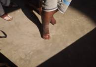
Case Report 1 A 62 year old man, a farmer with co-morbidities like bronchial asthma, inguinal hernia and a known smoker presented with an 8-10 weeks history of discharge and irregular, brownish, raised skin lesion over right dorsal aspect of foot. The patient did not have any history of trauma or insect bite. On examination a verrucose, brown to black, irregular swelling with discharge, about 2.2 cm X 4 cm X 1.5 cm, raised above skin surface was found. No offensive odour was noticed. No history of discharging granules was found. The discharge material collected aseptically for bacterial and fungal culture. A punch biopsy from the verrucose swelling was taken and put in 10% KOH solution and also inoculated aseptically for fungal culture. Subsequent examination of KOH preparation revealed presence of sclerotic bodies. Histologic opinion was also in favor of chromoblastomycosis without lymph adenopathy.
Figure1:Verrucous leg sweallings
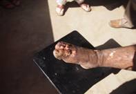
Case Report 2 A young (22 years) male tailor by profession, working in New Delhi, whose permanent address is Dalkhola, West Bengal, attended this skin OPD of the medical college on 16.07.2013 with the following complain of a brownish, raised, dry skin lesion over right thigh-6” above knee on inner aspect and a previously discharging similar lesion on right foot just above the little toe for about one year. He was referred to Microbiology dept. of this medical college for identification of fungal element from the lesions. On examination the lesions were crusty, verrucose, brownish, raised about 2cm above the skin surface, 1.5”-2” in size yielding a cauliflower like appearance. The lesion was confined to skin and subcutaneous tissue level. Initially the lesion began only on dorsum of right foot and after three months it started appearing on right medial aspect of thigh bellow right inguinal region. No enlarged inguinal lymph nodes were palpable on either side. KOH mount shows yellow brown bodies’ 6-8 µm in size having intersecting cross-walls. Histology report was in favor of Chromoblastomycosis.
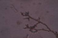
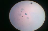
Figure 2: KOH preparation from culture – cladopsorium type Figure 3: KOH preparation shows medlar bodies
Discussion Chromoblastomycosis cases were mostly reported among rural population in countries like Madagascar in Africa and Brazil in South America. Few cases were also reported in India. In both the cases, the patients were immunocompetent. Under KOH preparations Sclerotic cells were noticed in both the cases from punch biopsy tissue. In India, Chromoblastomycosis has been reported from central Kerala among agricultural workers .No racial predilection was reported earlier but both the cases identified in our hospital were Muslims. No specific age group was related with the disease process. Chromoblastomycosis are very difficult to eradicate. Treatment with different antifungal drugs is tried but the results were not satisfactory. Recent antifungals with other treatment modalities were selected in our medical college with satisfactory results. According to Veraldi1 and Alan Coulson, only a few articles have been published on this topic over the last few decades. Veraldi also observed that only Candida as fungal pathogen was only identified in the leg ulcers with swelling in most of the patients in a wound care set up. As an exception we have identified unusual fungi in both the cases within a short span of six months in M.G.M. Medical College.
References
|
||||||
|
|||||||