 |
|||||||||||||||||||||||||||||||||||||||||||||||||||||||||||||||||||||||||||||||||||||||||||||||||||||||||||||||||||||||||||||||||||||||||||||||||||||||||||||||||||||||||||||||||||||||||||||||||||||||||||||||||||||||||||||||||||||||||||||
|
|
|||||||||||||||||||||||||||||||||||||||||||||||||||||||||||||||||||||||||||||||||||||||||||||||||||||||||||||||||||||||||||||||||||||||||||||||||||||||||||||||||||||||||||||||||||||||||||||||||||||||||||||||||||||||||||||||||||||||||||||
 |
|||||||||||||||||||||||||||||||||||||||||||||||||||||||||||||||||||||||||||||||||||||||||||||||||||||||||||||||||||||||||||||||||||||||||||||||||||||||||||||||||||||||||||||||||||||||||||||||||||||||||||||||||||||||||||||||||||||||||||||
|
|
[Abstract] [PDF] [HTML] [Linked References]
Clinical Profile and Prevalence of Opportunistic Infection in HIV Patients Attending Pediatric Department
Vikas N. Solunke1*, Milind B. Kamble2, Amol R. Suryawanshi3, Pallavi Saple4, Manish M. Tiwari5, Bhete S. B.6, Garad S. B.7 {1,3,5Assistant Professor, 4Professor and HOD, Department of Pediatrics} Swami Ramanand Teerth Rural Government Medical College and Hospital, Ambajogai, Maharashtra, INDIA. 2Professor and HOD, Department of Pediatrics, Shankarrao Chavan Government Medical College, Nanded, Maharashtra, INDIA. 6Department of Pharmacology, Topiwala National Medical College and Nayar Hospital, Mumbai, Maharashtra, INDIA. 7Medical Officer, Department of Ophthalmology, Government Medical College, Latur, Maharashtra, INDIA.*Corresponding Address: Research Article
Abstract: Introduction: It is important to concentrate on pediatric HIV as it differs from adult HIV regarding epidemiology, mode of transmission, diagnosis, immunology, pathology and clinical spectrum, management and presentation. The opportunistic infection also varies in pediatric and adult HIV regarding incidence, clinical manifestations, and presentations and after all its influence on morbidity and mortality. Aims and objectives: To study the clinical manifestations and prevalence of various opportunistic infections in pediatric HIV. Methodology: The present study was carried out in the department of pediatrics from January 2008 to June 2009. The study population included the patients who were already HIV positive or diagnosed later on investigation on suspicion of the clinical features, attending OPD or IPD of Pediatric department. Following criterion was used enroll the patients in the study. Total 144 HIV positive pediatric patients were diagnosed and were enrolled in the study. Detail history, clinical examination and lab investigation were conducted and various opportunistic infections were indentified in the study population. Results: It was observed that the majority (61.80%) cases were below 5 yrs of age. Severe malnutrition (Gr.III and IV) (59.02%) was the most common examination finding. It was followed by pallor (51.38%), respiratory signs (47.22%), lymphadenopathy (44.44%) etc. Prevalence of opportunistic infection in the present study was 83.3%. Among the opportunistic infections majority of cases were of tuberculosis (44.44%). It was followed by recurrent diarrhea (34.02%), recurrent pneumonia (29.86%), and oral candidiasis (25%) respectively. Conclusion: Prevalence of opportunistic infection in the present study was 83.3%.the most common opportunistic infection was TB followed by recurrent diarrhea and respiratory infection. Keywords: HIV Infection, pediatric.
Introduction Approximately 40 million people are living with HIV infection worldwide. This includes 2.5 million children with HIV infection and 0.7 million children are newly infected with HIV infection every year. There is a cumulative total of 11 million orphaned as their parent’s die of AIDS. In 2007about 420000 children <15 were newly infected with HIV1. Kaul D, Patel J2 described that in developed countries pediatric AIDS constitute only 2% of HIV infection whereas in developing countries it comprises about 20% of all the HIV infected cases due to greater affliction of women in child bearing age group. Impact of HIV/ AIDS in pediatrics age group is becoming one of the major and burning global problem affecting developing countries like India, which has a high prevalence and sharp rise with number of people living with HIV from few thousands in early 1995 to around 6 million adults and children in 2006. In India high prevalence is seen in Mumbai, Karnataka, and the Nagpur area of Maharashtra, Tamil Nadu, Coastal Andhra Pradesh and parts of Manipur and Nagaland in North East areas3. The increase in pediatric HIV infection has had a substantial impact on childhood mortality both in industrialized countries and developing countries4. As per Shivananda, Sanjeeva GN5 it is important to concentrate on pediatric HIV as it differs from adult HIV regarding epidemiology, mode of transmission, diagnosis, immunology, pathology and clinical spectrum, management and presentation. HIV infection result in gradual and progressive process that damage immune system of infected individual and make them susceptible to diverse collection of bacteria, virus, fungi, protozoa that represent the major cause of suffering and death for HIV infected person. These are opportunistic infections like Candida, Pneumocystis carnii, Cryptococcal meningitis, Herpes, Cytomegalo virus etc. affecting various organ system and fulminant group of disease, including severe pneumonia, TB, oropharyngeal candidiasis, chronic persistent diarrhoea, meningitis, encephalopathies, thrombocytopenic purpura, etc. The opportunistic infection also varies in pediatric and adult HIV regarding incidence, clinical manifestations, and presentations and after all its influence on morbidity and mortality. Hence it is essential to study OI’s in pediatric age group. This study in pediatric age group can help in early detection and finally some attempt to reduce the morbidity of patient and prolong the life span to some extent. Thus the present study was conducted to study various opportunistic infections occurring in pediatric HIV.
Aims and Objectives To study the clinical manifestations and prevalence of various opportunistic infections in pediatric HIV.Material and methods Study design: The present study was carried out in the department of pediatrics from January 2008 to June 2009. The study population included the patients who were already HIV positive or diagnosed later on investigation on suspicion of the clinical features, attending OPD or IPD of Pediatric department. Following criterion was used enroll the patients in the study. Inclusion criteria
A presumptive criterion of diagnosis of severe HIV disease
Other factors that support the diagnosis of severe HIV disease in the HIV sero positive infant include
In above condition confirmation of the diagnosis of HIV infection should be made as soon as possible. Exclusion criteria
Thus 144 were found to be HIV reactive.
Methodology Written and informed consent of all parents and care takers was taken before performing the tests and examination. Importance of test was explained to the parents. HIV was diagnosed by using following spots kits. 1. HIV comb (P.mittrs and Co. Pvt. Ltd., New Delhi), 2. Tri-Dot, 3. Callipus as rapid screening test and then ELISA were done. All those who are positive by ELISA were confirmed by repeat test after 3 months. All children who were thus confirmed HIV seropositive were included in this study. A detail history, physical examination, Anthropometric assessment and investigations was carried out as in all cases and entered on predesigned and pretested proforma. Laboratory evaluation was done in all cases for Hemoglobin, ESR, Total and Differential leucocyte count on peripheral blood smear (PS), Platelet count, Tuberculin test and Chest roentgenogram. If required the lymph node biopsy, CSF study, and blood culture and stool culture was done. Diagnosis of various opportunistic infection was done by using appropriate investigation where ever required. The data thus obtained was studied and the observations and results are tabulated and analyzed.
Precautions taken during investigations Full precautions were taken by using AIDS KIT containing disposable gown, face mask, cap, gloves, goggle, etc.
Results Table 1: Distribution of patients according to age, sex and area of residence
It was observed that the majority (61.80%) cases were below 5 yrs of age. Males (65.97%) outnumbered females (34.09%) with M: F ratio of 1.93:1. Maximum numbers of patients were from rural area (68.06%) whereas remaining were from urban area. Table 2: Distribution of patients according to various signs identified
In present study severe malnutrition (Gr.III and IV) (59.02%) was the most common examination finding. It was followed by pallor (51.38%), respiratory signs (47.22%), lymphadenopathy (44.44%), skin manifestations (33.33%), febrile (29.86%), hepatosplenomegaly (27.08), hepatomegaly (26.3). signs of vitamin deficiency (25%), Oral thrush (18.05%), CSOM (13.19%), splenomegaly (10.4%), Parotitis (8.33%) were the other observed signs.
Table 3: Distribution of patients according to various clinical symptoms on follow up
Out of the total 144 cases enrolled in the study, 100 cases had regular follow up for total 345 times and 44 cases didn’t come for follow up. Majority of follow ups were for respiratory complaints (22.89%) and fever (18.55%). Only 8.69% of asymptomatic patients came for follow up. Most of the follow up were for opportunistic infraction. Table 3: Distribution of patients according to various opportunistic infections
*Multiple responses were obtained
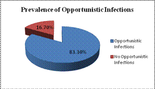
In the present study 144 cases were enrolled and studied for opportunistic infection if any. It was observed that only 24 cases had no evidence of any opportunistic infection. Whereas 124 cases had given evidence of opportunistic infection. Multiple opportunistic infections were observed in some cases. Thus prevalence of opportunistic infections in pediatric HIV patients was 83.3%.
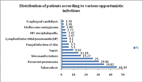
Among the opportunistic infections majority of cases were of tuberculosis (44.44%). It was followed by recurrent diarrhea (34.02%), recurrent pneumonia (29.86%), and oral candidiasis (25%) respectively. Pulmonary TB was found in majority of tuberculosis patient (56.25%) followed by extra-pulmonary TB (43.75%). Amongst 49 cases of recurrent diarrhea, in the majority of cases (55.1%) the cause could not be found out. But cryptosporidium was the most common single causative agent in rest of the cases where organisms were either cultured or found under microscopy.
Discussion The present study was conducted with the objective to study the clinical manifestations and prevalence of various opportunistic infections in pediatric HIV. In present study 61.80% children presented below 60 months (5 years) of age. The median age of presentation in this study was 4 yrs. The youngest patient being 3 months and oldest was 132 months. In the present series male outnumbered females with M: F ratio of 1.93:1. In present study maximum (68.05%) cases were from rural area and remaining was from urban area. Severe malnutrition in present series was observed in 92 (63.8%). Similar findings were also reported by Pol R et al6 in 54.9%, Emodi IJ, Okafor GO7 in 57% and Verghese V P et al8 in 58% cases. Merchant RH et al9 reported malnutrition only in 27% cases in their series. Malnutrition as a finding was seen in 100% cases by Lodha R et al10 and Diack et al11 in 89% which was very high figure as compared to present study and necessitating early nutritional intervention in HIV infected children. Cough was the second most common presenting complaint in the study and all of them showed some or the other respiratory finding. This was almost equal to the findings of Lodha R et al12. In the present study, skin rash were complained by 27.7% cases and on examination skin lesions were observed in 33.3% cases, thus emphasizing a role of detail clinical examination in HIV patients. The skin lesions were in the form disseminated scabies, pyoderma, pruritis, dermatitis, herpes, warts and molluscum contagiosum. The results observed in this study corroborates with that reported by Madhivanan P et al13. But the study from Delhi by Kaul D,Patel J14 reported low incidences as compared to present study, probably because this was retrospective case paper analysis, hence there might be under-reporting. Recently Pol R et al6 reports skin manifestation were more and more recognized and has been found in almost 60% pediatric HIV patients from Karnataka. In the present study pallor was a physical finding seen in 74(53.3%) cases which is higher than those reported by Pol R et al6 28.1%, Asnake S,Soloman A15 30.3% and Dhurat R et al16 32.4% Parthasarathy P et al17 40%. This high incidence of pallor might be due to association of malnutrition in 63% in the presents study. Out of the 144 cases during study period 120 had some or the other evidence of opportunistic infections hence the prevalence of OI was 83.3% in HIV infected children during study period. Sirisanthana V18 had reported prevalence of opportunistic infection in these children to be 79% which was almost equal to the present study. Gupta R et al19 reports the prevalence to be 65% in their study which was slightly lower than the present study, could be because of small sample size. TB has emerged as the most common opportunistic infection in the developing countries and was leader in opportunistic infections in the present series also (44.4% cases). Of the 64 cases of TB 36 cases were of pulmonary TB and 24 were of extrapulmonary TB. It is a known fact that HIV and TB co-exist in almost 22-48% children. TB has been reported by various authors from as low as 13.6% by Verghese P et al8 to as high as 70.9% by Asnake S,Soloman A15. Pol R et al6 showed TB in 38% of which pulmonary TB was present in a 59.3% and extrapulmonary in 40.7%. Such prevalence was also seen in the present study. Recurrent diarrhea was the 2nd most common opportunistic infection in present study accounting for 34.2% cases. Amongst the diarrheal cases cryptosporidium was culprit in 20.4% cases and in almost 55% the cause could not be found. These cases could be viral diarrheas. Recurrent diarrhea has been reported in 30.9% by Pol R et al6 and Verghese VP et al8 in 30% and Dhurat R et al16 in 27% cease which was comparable with the present study. Pneumonia in HIV is more prolonged and recurrent. Many times it is bacterial pneumonia in tropical and developing countries. Recurrent pneumonia in present series was seen in 29.8%cases. Madhivanan P et al13 has reported 36% cases with recurrent pneumonia and Dhurat R et al16 has reported 24.3%. Thus the result in present study was comparable. The prevalence of oral candidiasis was 25%; it was followed by skin manifestation in 15.27% cases and recurrent otits media in 13.19% cases. Sepsis was reported in 9.02% cases. Herpes zoster and fungal infection of skin was reported in 5.5% cases each. Warts, Lymphoid interstitial pneumonitis, Pneumocystitis carinii pneumonia, encephalopathy etc were also diagnosed in few cases but its prevalence was very less. Similar findings were also reported by Asnake S et al15, Merchant RH et al9, Madhivanan P et al13, Sirisanthana V18, Gupta R19 and Pol R et al6 with some variation.
Conclusion The prevalence of OI’s in HIV was 83.3%. TB was the most common opportunistic infection with prevalence of 44.4%. Followed by recurrent diarrhea (34.02%), recurrent pneumonia (29.86%) and oral candidiasis (25%) respectively. Therefore it can be concluded that with the rapid and continued spread of HIV pandemic and the increased survival of these victims, the opportunistic infections are on a rise.
References
|
||||||||||||||||||||||||||||||||||||||||||||||||||||||||||||||||||||||||||||||||||||||||||||||||||||||||||||||||||||||||||||||||||||||||||||||||||||||||||||||||||||||||||||||||||||||||||||||||||||||||||||||||||||||||||||||||||||||||||||
|
|||||||||||||||||||||||||||||||||||||||||||||||||||||||||||||||||||||||||||||||||||||||||||||||||||||||||||||||||||||||||||||||||||||||||||||||||||||||||||||||||||||||||||||||||||||||||||||||||||||||||||||||||||||||||||||||||||||||||||||

